. 2017 Mar 23:8:14890.
doi: 10.1038/ncomms14890.

Iron minerals within specific microfossil morphospecies of the 1.88 Ga Gunflint Formation

Affiliations

Iron minerals within specific microfossil morphospecies of the 1.88 Ga Gunflint Formation

Kevin Lepot et al. Nat Commun. .

Abstract

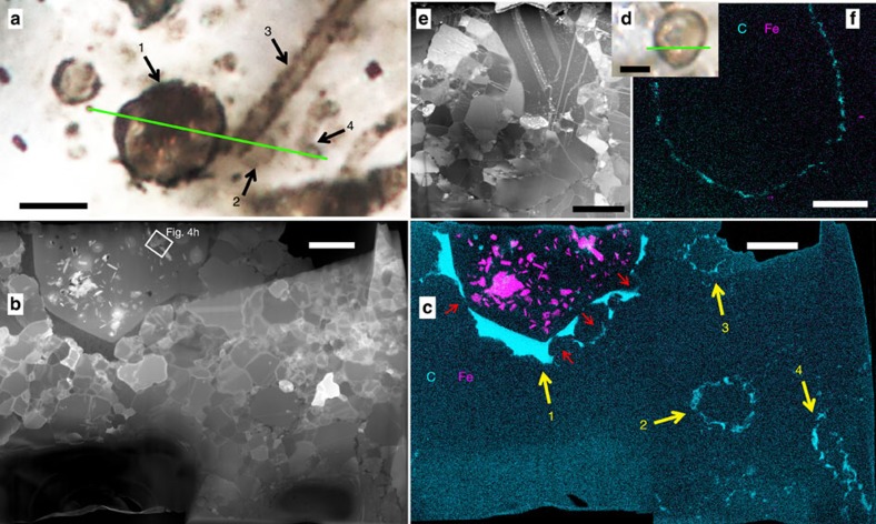
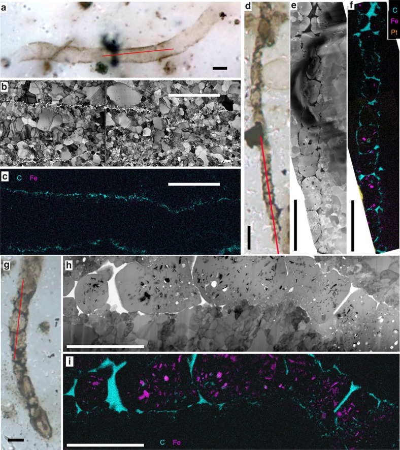
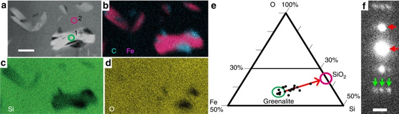
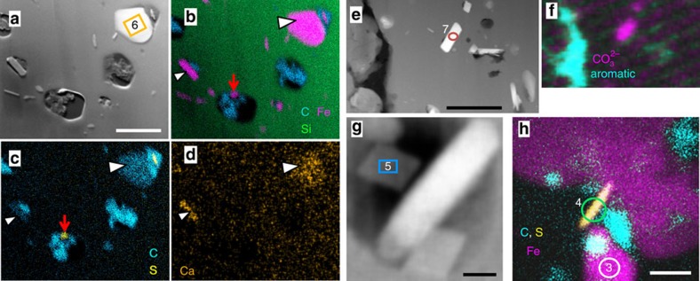
Problematic microfossils dominate the palaeontological record between the Great Oxidation Event 2.4 billion years ago (Ga) and the last Palaeoproterozoic iron formations, deposited 500-600 million years later. These fossils are often associated with iron-rich sedimentary rocks, but their affinities, metabolism, and, hence, their contributions to Earth surface oxidation and Fe deposition remain unknown. Here we show that specific microfossil populations of the 1.88 Ga Gunflint Iron Formation contain Fe-silicate and Fe-carbonate nanocrystal concentrations in cell interiors. Fe minerals are absent in/on all organically preserved cell walls. These features are consistent with in vivo intracellular Fe biomineralization, with subsequent in situ recrystallization, but contrast with known patterns of post-mortem Fe mineralization. The Gunflint populations that display relatively large cells (thick-walled spheres, filament-forming rods) and intra-microfossil Fe minerals are consistent with oxygenic photosynthesizers but not with other Fe-mineralizing microorganisms studied so far. Fe biomineralization may have protected oxygenic photosynthesizers against Fe2+ toxicity during the Palaeoproterozoic.


Conflict of interest statement

The authors declare no competing financial interests.

Figures

Figure 1
Figure 1. Iron in Gunflint microfossils.
(ac) 1: thick-walled Huroniospora, 2: thin-walled Huroniospora, 3–4: Type 1 (cell-free sheaths) G. minuta. (df) Thin-walled Huroniospora. (a,d) Multiplane photomicrographs. Scale bars, 5 μm. (b,e) STEM dark-field images of the FIB ultrathin sections cut along the green lines. Scale bars, 2 μm. (c,f) STEM maps of Fe (pink), C (cyan); corresponding maps of Si, O and S are shown in Supplementary Fig. 4. Scale bars, 2 μm. Fe minerals are highly concentrated inside thick-walled Huroniospora and nearly absent in or near Type 1 G. minuta and thin-walled Huroniospora. Red arrows in c indicate displacement of wall organic matter by quartz grains.
Figure 2
Figure 2. Iron in Gunflint filaments.
Scale bars, 5 μm. (ac) Animikiea of Type 1 (without cell remnants in sheath). (df) G. minuta of Type 2 (with cell-like segmentation). (gi) Type 2 G. grandis. (a,d,g) Multiplane photomicrographs. (b,e,h) STEM bright- (b,h) and dark- (e) field images of the FIB ultrathin sections cut along the red lines. (c,f,i) STEM maps of Fe (pink), C (cyan) and preparation coatings (Pt: orange); corresponding maps of Si, O and S are shown in Supplementary Fig. 7. Fe minerals occur in G. grandis and G. minuta Type 2, but not in Animikiea. Fe minerals are absent in the vicinity of these microfossils.
Figure 3
Figure 3. Intra-microfossil greenalite.
(ad) STEM of greenalite in quartz. (a) Dark-field image. Scale bar, 200 nm. Circles numbered 1 and 2 outline target areas of EDXS and EELS spectra (displayed in Supplementary Fig. 15 with corresponding numbers). (bd) EDXS mappings of Fe (pink in b), organic C (cyan in b), Si (green in c), O (yellow in d) in the region in a. The crystals in the green circle traverse the FIB section, allowing EDXS analyses without quartz interference, as confirmed by Si EELS spectra. (e) Ternary Fe–Si–O (atomic %) plot derived from EDXS spectra (Supplementary Fig. 15c): quartz-free greenalites (including that numbered 1 in a) plot in the green circle, whereas other crystals plot on a mixing line with quartz (pink circle), indicating that all greenalite crystals have a similar composition. The Si/O ratio of 0.32 (n=4 quartz-free crystals) is consistent with the general formula of greenalite: Si2O5(OH)4(Fe2+,Fe3+)2–3. EELS spectra display a Fe3+/Fetotal <4.5‰ (Supplementary Fig. 15e), indicating a composition close to Si2O5(OH)4(Fe2+)3. (f) SAED pattern recorded on the region numbered 1 in a. Scale bar, 1 nm−1. Arrows indicate lattice spacing diagnostic of greenalite: 7.2 Å (0,0,1) planes (red), and 23 Å superlattice (green).
Figure 4
Figure 4. Intra-microfossil siderite and Fe2+ sulfides.
(a) STEM dark-field image. (bd) EDXS mappings of a showing quartz (Si: green in b), siderite (Fe: pink in b, minor Ca: orange in d, arrowheads), and Fe+S minerals (S in yellow in c, red arrows in b,c) that are systematically nanoscale and embedded in organic matter (C: cyan in b,c). Scale bar, 500 nm. (e,f) STEM dark-field image (e) and STXM mapping (f) of e showing carbonate (pink) and aromatic carbon (cyan). The rod-shaped (red circle) crystal is siderite. Scale bar, 500 nm. (g) STEM dark-field image showing rhombohedra of siderite (for example, blue box) associated with greenalite (white rod). Scale bar, 50 nm. (h) EDXS mapping of a nanoscale Fe2+ and S (yellow) crystal embedded in organic carbon (cyan) between two greenalite (Fe: pink) crystals. Scale bar, 100 nm. Numbered boxes and circles in a,e,g,h outline target areas of EDXS spectra displayed in Supplementary Figs 15 and 16 with corresponding numbers.

References

    1. Planavsky N. et al.. Iron isotope composition of some Archean and Proterozoic iron formations. Geochim. Cosmochim. Acta 80, 158–169 (2012).
    1. Lyons T. W., Reinhard C. T. & Planavsky N. J. The rise of oxygen in Earth's early ocean and atmosphere. Nature 506, 307–315 (2014). - PubMed
    1. Rasmussen B. et al.. Deposition of 1.88-billion-year-old iron formations as a consequence of rapid crustal growth. Nature 484, 498–501 (2012). - PubMed
    1. Fortin D. & Langley S. Formation and occurrence of biogenic iron-rich minerals. Earth Sci. Rev. 72, 1–19 (2005).
    1. Klueglein N. et al.. Potential role of nitrite for abiotic Fe(II) oxidation and cell encrustation during nitrate reduction by denitrifying bacteria. Appl. Environ. Microbiol. 80, 1051–1061 (2014). - PMC - PubMed

Publication types