

Profiling protein expression in circulating tumour cells using microfluidic western blotting
- PMID: 28332571
- PMCID: PMC5376644
- DOI: 10.1038/ncomms14622
Profiling protein expression in circulating tumour cells using microfluidic western blotting
Abstract
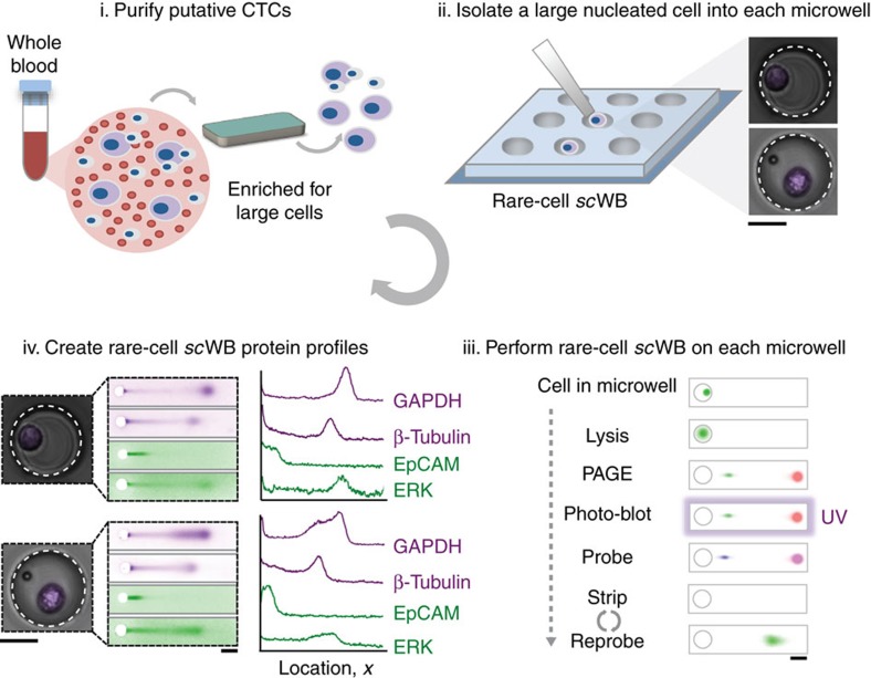
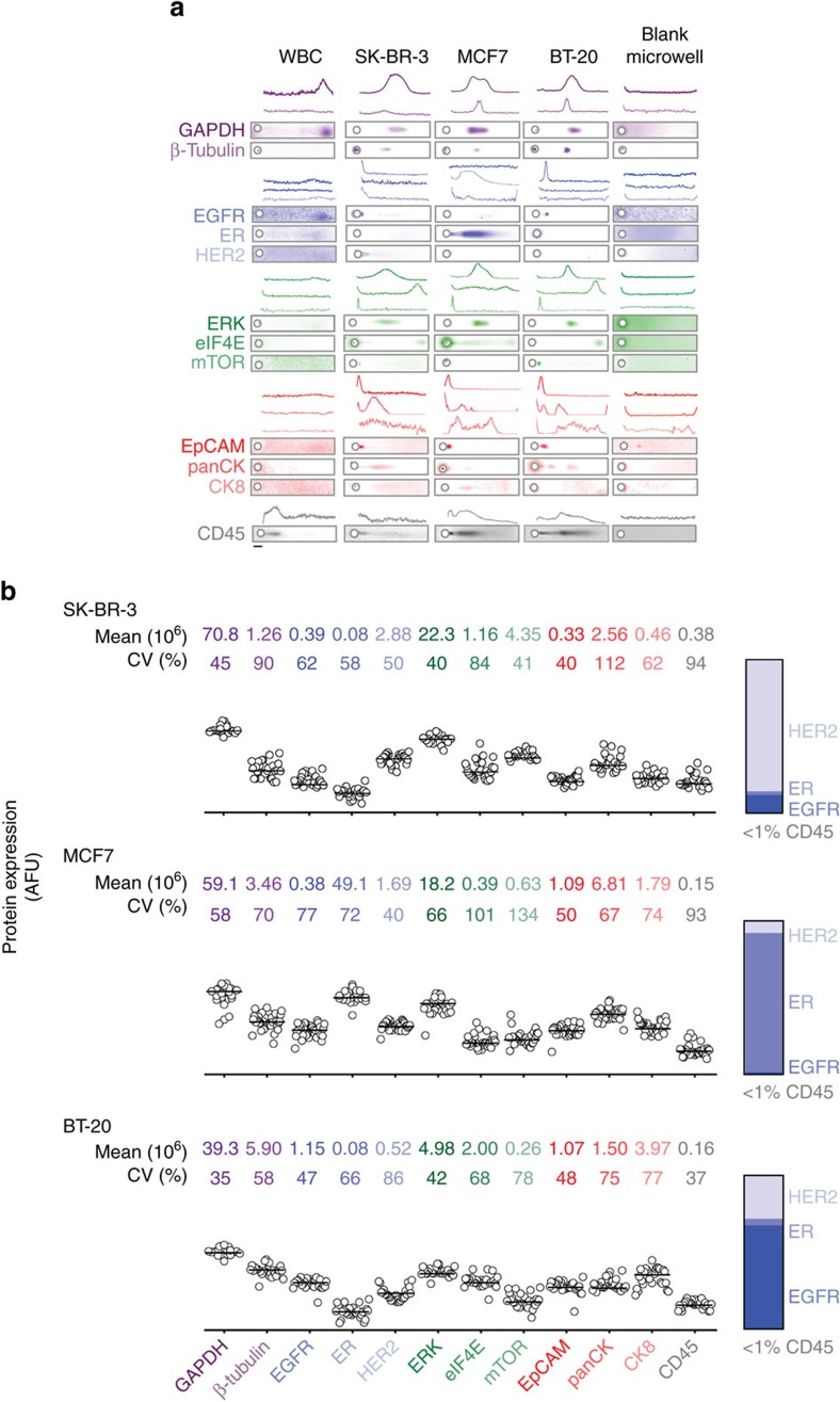
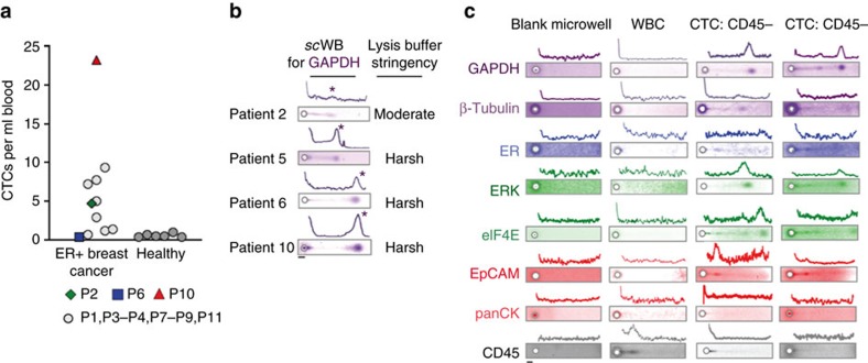
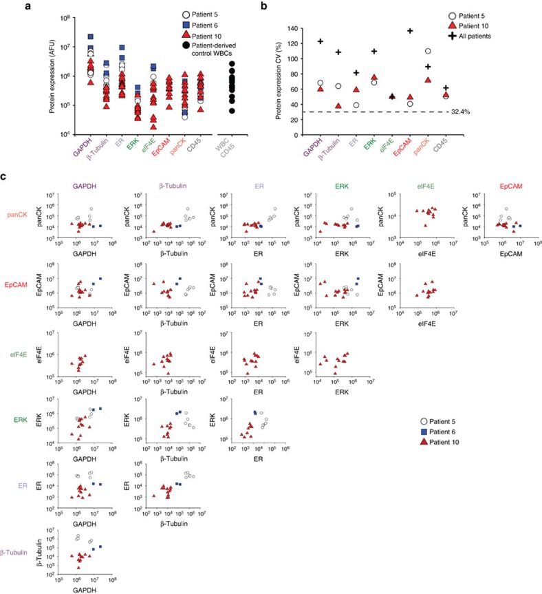
Circulating tumour cells (CTCs) are rare tumour cells found in the circulatory system of certain cancer patients. The clinical and functional significance of CTCs is still under investigation. Protein profiling of CTCs would complement the recent advances in enumeration, transcriptomic and genomic characterization of these rare cells and help define their characteristics. Here we describe a microfluidic western blot for an eight-plex protein panel for individual CTCs derived from estrogen receptor-positive (ER+) breast cancer patients. The precision handling and analysis reveals a capacity to assay sparingly available patient-derived CTCs, a biophysical CTC phenotype more lysis-resistant than breast cancer cell lines, a capacity to report protein expression on a per CTC basis and two statistically distinct GAPDH subpopulations within the patient-derived CTCs. Targeted single-CTC proteomics with the capacity for archivable, multiplexed protein analysis offers a unique, complementary taxonomy for understanding CTC biology and ascertaining clinical impact.
Conflict of interest statement
E.S., T.A.D., J.V., K.A.Y. and A.E.H. has financial interest in intellectual property related to the device and assay described here, and may benefit from royalties from licensing. Further, A.E.H. has financial interest in commercialization efforts. E.S.C., C.R. and J.C. have financial interests in Vortex Biosciences. S.S.J. has research funded by Vortex Biosciences. All other authors declare no competing financial interest.
Figures
Similar articles
-
An integrated microfluidic chip system for single-cell secretion profiling of rare circulating tumor cells.Sci Rep. 2014 Dec 16;4:7499. doi: 10.1038/srep07499. Sci Rep. 2014. PMID: 25511131 Free PMC article.
-
Single cell profiling of circulating tumor cells: transcriptional heterogeneity and diversity from breast cancer cell lines.PLoS One. 2012;7(5):e33788. doi: 10.1371/journal.pone.0033788. Epub 2012 May 7. PLoS One. 2012. PMID: 22586443 Free PMC article.
-
Correlation of hormone receptor status between circulating tumor cells, primary tumor, and metastasis in breast cancer patients.Clin Transl Oncol. 2015 Jul;17(7):539-46. doi: 10.1007/s12094-015-1275-1. Epub 2015 Jan 23. Clin Transl Oncol. 2015. PMID: 25613123 Free PMC article.
-
Microfluidics and circulating tumor cells.J Mol Diagn. 2013 Mar;15(2):149-57. doi: 10.1016/j.jmoldx.2012.09.004. Epub 2012 Dec 22. J Mol Diagn. 2013. PMID: 23266318 Review.
-
Circulating tumor cells in breast cancer: applications in personalized medicine.Breast Cancer Res Treat. 2016 Dec;160(3):411-424. doi: 10.1007/s10549-016-4014-6. Epub 2016 Oct 19. Breast Cancer Res Treat. 2016. PMID: 27761678 Review.
Cited by
-
Biomedical Applications of Microfluidic Devices: A Review.Biosensors (Basel). 2022 Nov 16;12(11):1023. doi: 10.3390/bios12111023. Biosensors (Basel). 2022. PMID: 36421141 Free PMC article. Review.
-
Microparticle Delivery of Protein Markers for Single-Cell Western Blotting from Microwells.Small. 2018 Nov;14(48):e1802865. doi: 10.1002/smll.201802865. Epub 2018 Oct 17. Small. 2018. PMID: 30334351 Free PMC article.
-
Deep Profiling of Cellular Heterogeneity by Emerging Single-Cell Proteomic Technologies.Proteomics. 2020 Jul;20(13):e1900226. doi: 10.1002/pmic.201900226. Epub 2019 Dec 2. Proteomics. 2020. PMID: 31729152 Free PMC article. Review.
-
Protein fishing from single live cells.J Nanobiotechnology. 2018 Sep 11;16(1):67. doi: 10.1186/s12951-018-0395-5. J Nanobiotechnology. 2018. PMID: 30205820 Free PMC article.
-
Versatile Microfluidic Mixing Platform for High- and Low-Viscosity Liquids via Acoustic and Chemical Microbubbles.Micromachines (Basel). 2019 Dec 5;10(12):854. doi: 10.3390/mi10120854. Micromachines (Basel). 2019. PMID: 31817508 Free PMC article.
References
-
- Sollier E. et al.. Size-selective collection of circulating tumor cells using Vortex technology. Lab Chip 14, 63–77 (2014). - PubMed
-
- Zheng S. et al.. Membrane microfilter device for selective capture, electrolysis and genomic analysis of human circulating tumor cells. J. Chromatogr. A 1162, 154–161 (2007). - PubMed
Publication types
MeSH terms
Substances
Grants and funding
LinkOut - more resources
Full Text Sources
Other Literature Sources
Medical
Research Materials
