. 2017 Mar 23:7:45146.
doi: 10.1038/srep45146.

Xeno-free culture of human pluripotent stem cells on oligopeptide-grafted hydrogels with various molecular designs

Affiliations

Xeno-free culture of human pluripotent stem cells on oligopeptide-grafted hydrogels with various molecular designs

Yen-Ming Chen et al. Sci Rep. .

Abstract

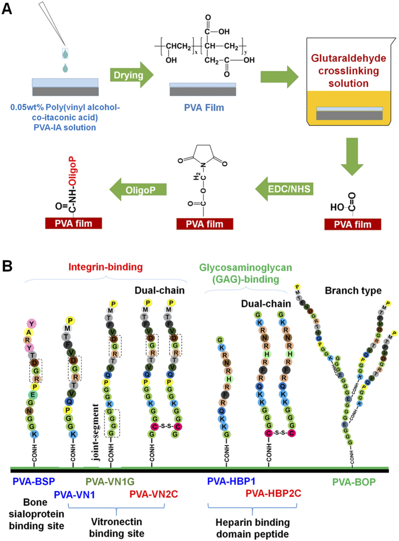
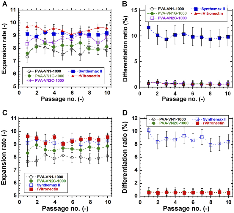
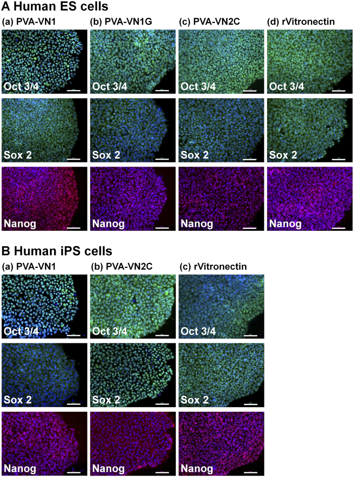
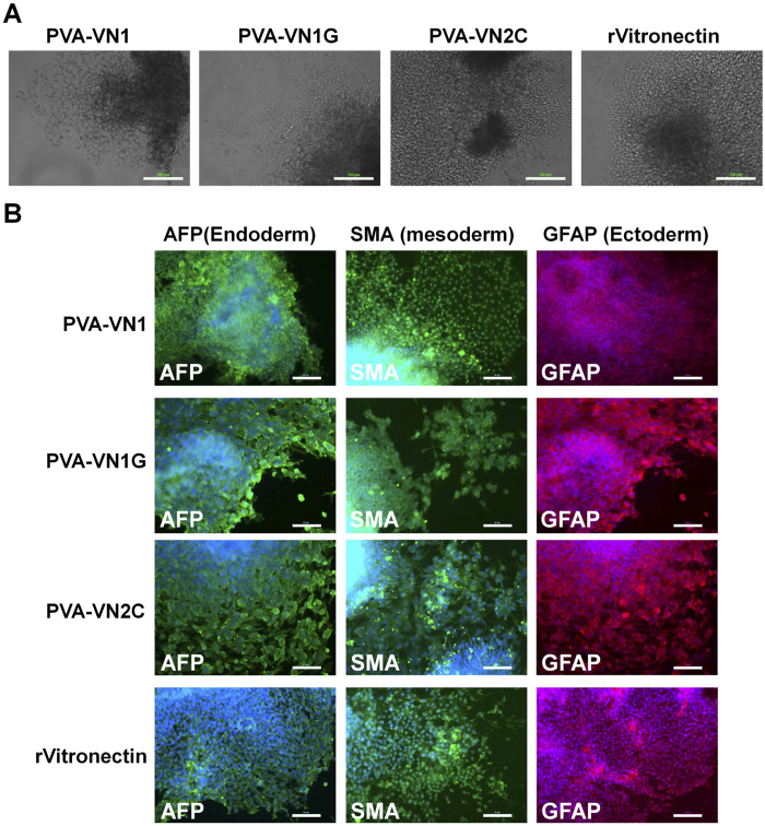
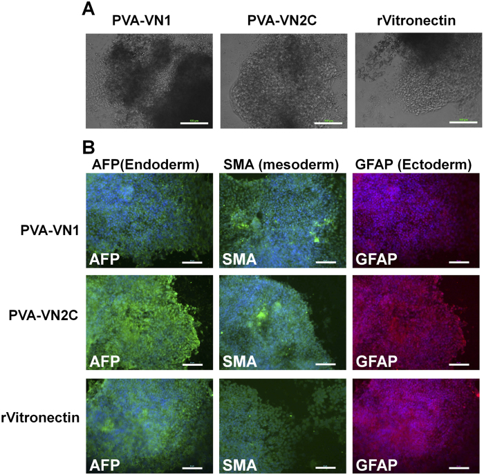
Establishing cultures of human embryonic (ES) and induced pluripotent (iPS) stem cells in xeno-free conditions is essential for producing clinical-grade cells. Development of cell culture biomaterials for human ES and iPS cells is critical for this purpose. We designed several structures of oligopeptide-grafted poly (vinyl alcohol-co-itaconic acid) hydrogels with optimal elasticity, and prepared them in formations of single chain, single chain with joint segment, dual chain with joint segment, and branched-type chain. Oligopeptide sequences were selected from integrin- and glycosaminoglycan-binding domains of the extracellular matrix. The hydrogels grafted with vitronectin-derived oligopeptides having a joint segment or a dual chain, which has a storage modulus of 25 kPa, supported the long-term culture of human ES and iPS cells for over 10 passages. The dual chain and/or joint segment with cell adhesion molecules on the hydrogels facilitated the proliferation and pluripotency of human ES and iPS cells.


Conflict of interest statement

The authors declare no competing financial interests.

Figures

Figure 1
Figure 1. Preparation and design of PVA hydrogels grafted with several oligopeptides containing cell-binding domains.
(A) A chemical scheme for PVA hydrogels reacting with oligopeptides (oligoP). (B) Design and sequence of oligopeptides grafted on PVA hydrogels. Single chains (PVA-BSP, PVA-VN1, and PVA-HBP1), single chains with a joint segment (PVA-VN1G), dual chains (PVA-VN2C and PVA-HBP2C), and branch-type (PVA-BOP) oligopeptides were grafted on PVA hydrogels. PVA-BSP, PVA-VN1, PVA-VN1G, PVA-VN2C, and PVA-BOP have an integrin-binding domain, whereas PVA-HBP1 and PVA-HBP2C have a glycosaminoglycan-binding domain.
Figure 2
Figure 2. Characterization of PVA hydrogels grafted with various oligopeptides.
(A) High-resolution XPS spectra of the C1s peaks analyzed on the surface of unmodified PVA (a), PVA-VN1-1000 (b), PVA-VN1G-1000 (c), and PVA-VN2C-1000 (d) hydrogels. (B) High-resolution XPS spectra of the N1s peaks analyzed on the surface of unmodified PVA (a), PVA-VN1-1000 (b), PVA-VN1G-1000 (c), and PVA-VN2C-1000 (d) hydrogels. (C) The nitrogen to carbon (N/C) atomic ratios in PVA and PVA-BSP, PVA-VN1, PVA-VN1G, PVA-VN2C, PVA-BOP, PVA-HBP1, and PVA-HBP2C hydrogels grafted with different concentrations of oligopeptides (200, 500, or 1000 μg/mL).
Figure 3
Figure 3. Human ES cells cultured on PVA hydrogels grafted with different design of oligopeptides under xeno-free culture conditions.
(A) Morphology of human ES (H9) cells cultivated on PVA hydrogels grafted with various oligopeptides (PVA-BSP-1000, PVA-VN1-1000, PVA-VN1G-1000, PVA-VN2C-1000, PVA-BOP-100, PVA-HBP1-1000, and PVA-HBP2C-1000) and on Synthemax II-coated dishes at passage 1. Scale bar indicates 100 μm. (B) Average doubling time of human ES (H9) cells on PVA hydrogels grafted with various oligopeptides (PVA-BSP-500, PVA-BSP-1000, PVA-VN1-500, PVA-VN1-1000, PVA-VN1G-500, PVA-VN1G-1000, PVA-VN2C-500, PVA-VN2C-1000, PVA-BOP-10, PVA-BOP-50, and PVA-BOP-100), Synthemax II-coated dishes, and rVitronectin-coated dishes during passages 1–3. *p < 0.05; **p < 0.05; ***p < 0.05. (C) Differentiation ratio of human ES (H9) cells on PVA hydrogels grafted with various oligopeptides (PVA-BSP-500, PVA-BSP-1000, PVA-VN1-500, PVA-VN1-1000, PVA-VN1G-500, PVA-VN1G-1000, PVA-VN2C-500, PVA-VN2C-1000, PVA-BOP-10, PVA-BOP-50, and PVA-BOP-100), Synthemax II-coated dishes, and rVitronectin-coated dishes during passages 1–3.
Figure 4
Figure 4. Human ES cells cultured on PVA hydrogels grafted with different design of oligopeptides under xeno-free culture conditions, prepared with a low oligopeptide concentration (200 μg/mL).
(A) Morphology of human ES (H9) cells cultivated on PVA hydrogels grafted with various oligopeptides (a) PVA-BSP-200, (b) PVA-VN1-200, (c) PVA-VN1G-200, and (d) PVA-VN2C-200), on (e) Synthemax II-coated dishes, and on (f) rVitronectin-coated dishes at passage 1. Scale bar indicates 500 μm. (B) Average doubling time of human ES (H9) cells grown on PVA hydrogels grafted with various oligopeptides (PVA-BSP-200, PVA-VN1-200, PVA-VN1G-200, PVA-VN2C-200, and PVA-VN2C-1000), on Synthemax II-coated dishes, and on rVitronectin-coated dishes during passages 1–3. (C) Differentiation ratio of human ES (H9) cells grown on PVA hydrogels grafted with various oligopeptides (PVA-BSP-200, PVA-VN1-200, PVA-VN1G-200, PVA-VN2C-200, and PVA-VN2C-1000), on Synthemax II-coated dishes, and on rVitronectin-coated dishes during passages 1–3.
Figure 5
Figure 5. Long-term culture of human ES and iPS cells on PVA hydrogels grafted with different oligopeptide designs under xeno-free culture conditions.
(A) Expansion rate and (B) differentiation ratio of human ES (H9) cells on PVA-VN1-1000 hydrogels (open black circle), PVA-VN1G-1000 hydrogels (closed green circle), PVA-VN2C-1000 hydrogels (open purple square), Synthemax II-coated dishes (closed blue square), and rVitronectin-coated dishes (closed red triangle) for 10 passages. (C) Expansion rate and (D) differentiation ratio of human iPS (HPS0077) cells on PVA-VN1-1000 hydrogels (open black circle), PVA-VN2C-1000 hydrogels (closed green circle), Synthemax II-coated dishes (open blue square), and rVitronectin-coated dishes (closed red square) for 10 passages.
Figure 6
Figure 6. Characterization of the pluripotency of human ES and iPS cells when cultured on PVA hydrogels grafted with different oligopeptide designs under xeno-free culture conditions for 10 passages.
(A) Expression of pluripotency proteins Oct3/4 (green), Sox2 (green), and Nanog (red) on human ES (H9) cells evaluated by immunostaining with dual staining with Hoechest33342 for nuclear (blue) after culturing on (a) PVA-VN1-1000, (b) PVA-VN1G-1000, and (c) PVA-VN2C-1000 hydrogels, and on (d) rVitronectin-coated dishes under xeno-free conditions for 10 passages. (B) Expression of pluripotency proteins Oct3/4 (green), Sox2 (green), and Nanog (red) on human iPS (HPS0077) cells evaluated by immunostaining with dual staining with Hoechest33342 for nuclear (blue) after culturing on (a) PVA-VN1-1000 and (b) PVA-VN2C-1000 hydrogels and on (c) rVitronectin-coated dishes under xeno-free conditions for 10 passages. Scale bar indicates 50 μm.
Figure 7
Figure 7. Characterization of the differentiation ability of human ES cells when cultured on PVA hydrogels grafted with different oligopeptide designs under xeno-free culture conditions for 10 passages.
(A) Morphology of cells from EBs differentiated from human ES (H9) cells after culturing on (a) PVA-VN1-1000, (b) PVA-VN1G-1000, (c) PVA-VN2C-1000 hydrogels, and (d) rVitronectin-coated dishes under xeno-free conditions for 10 passages. Scale bar indicates 100 μm. (B) Expression of an endoderm protein (AFP, green), mesoderm protein (SMA, green), and ectoderm protein (GFAP, red) in human ES (H9) cells evaluated by immunostaining with dual staining with Hoechest33342 for nuclear (blue) after culturing on PVA-VN1-1000, PVA-VN1G-1000, and PVA-VN2C-1000 hydrogels, and on (d) rVitronectin-coated dishes under xeno-free conditions for 10 passages. Scale bar indicates 50 μm.
Figure 8
Figure 8. Characterization of the differentiation ability of human iPS cells when cultured on PVA hydrogels grafted with different oligopeptide designs under xeno-free culture conditions after culturing for 10 passages.
(A) Morphology of cells from EBs differentiated from human iPS (HPS0077) cells after culturing on PVA-VN1-1000 and PVA-VN2C-1000 hydrogels, and on rVitronectin-coated dishes under xeno-free conditions for 10 passages. Scale bar indicates 100 μm. (B) Expression of an endoderm protein (AFP, green), mesoderm protein (SMA, green), and ectoderm protein (GFAP, red) in human iPS (HPS0077) cells evaluated by immunostaining with dual staining with Hoechest33342 for nuclear (blue) after culturing on PVA-VN1-1000 and PVA-VN2C-1000 hydrogels, and on rVitronectin-coated dishes under xeno-free conditions for 10 passages. Scale bar indicates 50 μm.
Figure 9
Figure 9. Characterization of the differentiation ability of human ES (H9) cells in vivo after culturing on PVA-VN1-1000 and PVA-VN2C hydrogels under xeno-free cell culture conditions for 10 passages.
(A) (a) A picture of teratoma by injecting with human ES cells after culturing on PVA-VN1-1000 hydrogels after 10 passages. Tissues including (b) intestinal epithelium (endoderm), (c) cartilage (mesoderm), and (d) neuroepithelium (ectoderm) can be observed. (B) (a) A picture of teratoma by injecting with human ES cells after culturing on PVA-VN2C-1000 hydrogels after 10 passages. Tissues including (b) intestinal epithelium (endoderm), (c) cartilage (mesoderm), and (d) retinal pigment epithelium (ectoderm) can be observed.

References

    1. Higuchi A. et al.. Biomaterials for the feeder-free culture of human embryonic stem cells and induced pluripotent stem cells. Chem. Rev. 111, 3021–3035 (2009). - PubMed
    1. Higuchi A. et al.. Design of polymeric materials for culturing human pluripotent stem cells: Progress toward feeder-free and xeno-free culturing. Prog. Polym. Sci. 39, 1348–1374 (2014).
    1. Takahashi K. et al.. Induction of pluripotent stem cells from adult human fibroblasts by defined factors. Cell. 131, 861–872 (2007). - PubMed
    1. Yu J. et al.. Induced pluripotent stem cell lines derived from human somatic cells. Science 318, 1917–1920 (2007). - PubMed
    1. Thomson J. A. et al.. Embryonic stem cell lines derived from human blastocysts. Science 282, 1145–1147 (1998). - PubMed

Publication types

MeSH terms