

Long-term white matter tract reorganization following prolonged febrile seizures
- PMID: 28332711
- PMCID: PMC5484997
- DOI: 10.1111/epi.13724
Long-term white matter tract reorganization following prolonged febrile seizures
Abstract
Objective: Diffusion magnetic resonance imaging (MRI) studies have demonstrated acute white matter changes following prolonged febrile seizures (PFS), but their longer-term evolution is unknown. We investigated a population-based cohort to determine white matter diffusion properties 8 years after PFS.
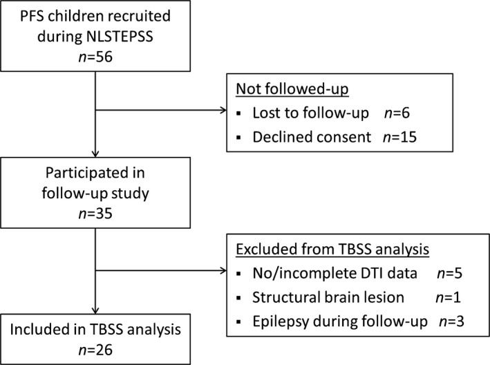
Methods: We used diffusion tensor imaging (DTI) and applied Tract-Based Spatial Statistics for voxel-wise comparison of white matter microstructure between 26 children with PFS and 27 age-matched healthy controls. Age, gender, handedness, and hippocampal volumes were entered as covariates for voxel-wise analysis.
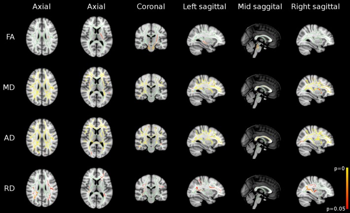
Results: Mean duration between the episode of PFS and follow-up was 8.2 years (range 6.7-9.6). All children were neurologically normal, and had normal conventional neuroimaging. On voxel-wise analysis, compared to controls, the PFS group had (1) increased fractional anisotropy in early maturing central white matter tracts, (2) increased mean and axial diffusivity in several peripheral white matter tracts and late-maturing central white matter tracts, and (3) increased radial diffusivity in peripheral white matter tracts. None of the tracts had reduced fractional anisotropy or diffusivity indices in the PFS group.
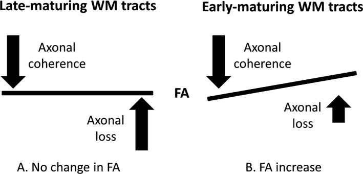
Significance: In this homogeneous, population-based sample, we found increased fractional anisotropy in early maturing central white matter tracts and increased mean and axial diffusivity with/without increased radial diffusivity in several late-maturing peripheral white matter tracts 8 years post-PFS. We propose disruption in white matter maturation secondary to seizure-induced axonal injury, with subsequent neuroplasticity and microstructural reorganization as a plausible explanation.
Keywords: Axonal injury; Diffusion tensor imaging; Neuroplasticity; Status epilepticus; White matter.
© 2017 The Authors. Epilepsia published by Wiley Periodicals, Inc. on behalf of International League Against Epilepsy.
Figures
Similar articles
-
Altered White Matter Microstructure Correlates with IQ and Processing Speed in Children and Adolescents Post-Fontan.J Pediatr. 2018 Sep;200:140-149.e4. doi: 10.1016/j.jpeds.2018.04.022. Epub 2018 Jun 19. J Pediatr. 2018. PMID: 29934026
-
Diffusion tensor imaging in children with tuberous sclerosis complex: tract-based spatial statistics assessment of brain microstructural changes.Pediatr Radiol. 2016 Jul;46(8):1158-64. doi: 10.1007/s00247-016-3582-2. Epub 2016 Apr 16. Pediatr Radiol. 2016. PMID: 27085522
-
Prolonged febrile seizures cause reversible reductions in white matter integrity.Neuroimage Clin. 2013 Oct 24;3:515-21. doi: 10.1016/j.nicl.2013.10.010. eCollection 2013. Neuroimage Clin. 2013. PMID: 24273734 Free PMC article.
-
Hemispheric Regional Based Analysis of Diffusion Tensor Imaging and Diffusion Tensor Tractography in Patients with Temporal Lobe Epilepsy and Correlation with Patient outcomes.Sci Rep. 2019 Jan 18;9(1):215. doi: 10.1038/s41598-018-36818-x. Sci Rep. 2019. PMID: 30659215 Free PMC article.
-
Diffusion-based magnetic resonance imaging and tractography in epilepsy.Epilepsia. 2008 Feb;49(2):189-200. doi: 10.1111/j.1528-1167.2007.01378.x. Epub 2007 Oct 16. Epilepsia. 2008. PMID: 17941849 Review.
Cited by
-
Febrile seizures: an overview.Drugs Context. 2018 Jul 16;7:212536. doi: 10.7573/dic.212536. eCollection 2018. Drugs Context. 2018. PMID: 30038660 Free PMC article. Review.
-
Alterations of Hippocampal Myelin Sheath and Axon Sprouting by Status Convulsion and Regulating Lingo-1 Expression with RNA Interference in Immature and Adult Rats.Neurochem Res. 2018 Mar;43(3):721-735. doi: 10.1007/s11064-018-2474-2. Epub 2018 Jan 27. Neurochem Res. 2018. PMID: 29383653
-
Subcortical nuclei volumes are associated with cognition in children post-convulsive status epilepticus: Results at nine years follow-up.Epilepsy Behav. 2020 Sep;110:107119. doi: 10.1016/j.yebeh.2020.107119. Epub 2020 Jun 8. Epilepsy Behav. 2020. PMID: 32526686 Free PMC article.
-
Transcranial magnetic stimulation (TMS) seeded tractography provides superior prediction of eloquence compared to anatomic seeded tractography.Neurooncol Adv. 2022 Sep 15;4(1):vdac126. doi: 10.1093/noajnl/vdac126. eCollection 2022 Jan-Dec. Neurooncol Adv. 2022. PMID: 36128584 Free PMC article.
-
Exploring Variances of White Matter Integrity and the Glymphatic System in Simple Febrile Seizures and Epilepsy.Front Neurol. 2021 Apr 21;12:595647. doi: 10.3389/fneur.2021.595647. eCollection 2021. Front Neurol. 2021. PMID: 33967932 Free PMC article.
References
-
- Chin RF, Neville BG, Peckham C, et al. Incidence, cause, and short‐term outcome of convulsive status epilepticus in childhood: prospective population‐based study. Lancet 2006;368:222–229. - PubMed
-
- Provenzale JM, Barboriak DP, VanLandingham K, et al. Hippocampal MRI signal hyperintensity after febrile status epilepticus is predictive of subsequent mesial temporal sclerosis. AJR Am J Roentgenol 2008;190:976–983. - PubMed
-
- Scott RC, King MD, Gadian DG, et al. Hippocampal abnormalities after prolonged febrile convulsion: a longitudinal MRI study. Brain 2003;126:2551–2557. - PubMed
-
- Tarkka R, Paakko E, Pyhtinen J, et al. Febrile seizures and mesial temporal sclerosis: no association in a long‐term follow‐up study. Neurology 2003;60:215–218. - PubMed
Publication types
MeSH terms
Grants and funding
LinkOut - more resources
Full Text Sources
Other Literature Sources
