

The intravoxel incoherent motion MRI of lateral pterygoid muscle: a quantitative analysis in patients with temporomandibular joint disorders
- PMID: 28332854
- PMCID: PMC5595037
- DOI: 10.1259/dmfr.20160424
The intravoxel incoherent motion MRI of lateral pterygoid muscle: a quantitative analysis in patients with temporomandibular joint disorders
Abstract
Objectives: To quantitatively evaluate diffusion and perfusion status of lateral pterygoid muscle (LPM) in patients with temporomandibular joint disorder (TMD) by intravoxel incoherent motion (IVIM) imaging and to correlate with findings on temporomandibular joints (TMJs) by conventional MRI.
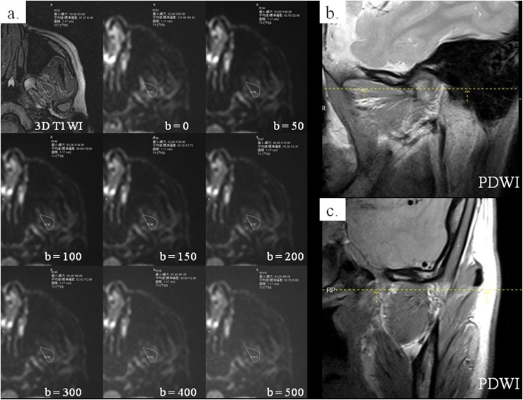
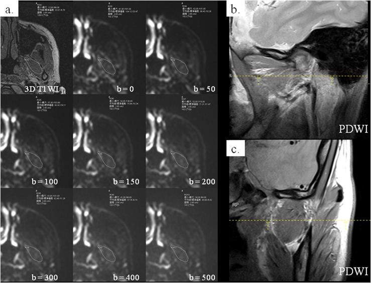
Methods: 42 patients with TMD underwent MRI. To assess IVIM parameters, diffusion-weighted imaging was obtained by spin-echo-based single-shot echoplanar imaging. Regions of interest were created on all diffusion-weighted images of the superior belly of the lateral pterygoid (SLP) and inferior belly of the lateral pterygoid (ILP) at b-values 0-500 s mm-2. Then, IVIM parameters, diffusion (D) and perfusion (f) were calculated using biexponential fittings. The correlation of these values with conventional MRI findings on TMJs was investigated.
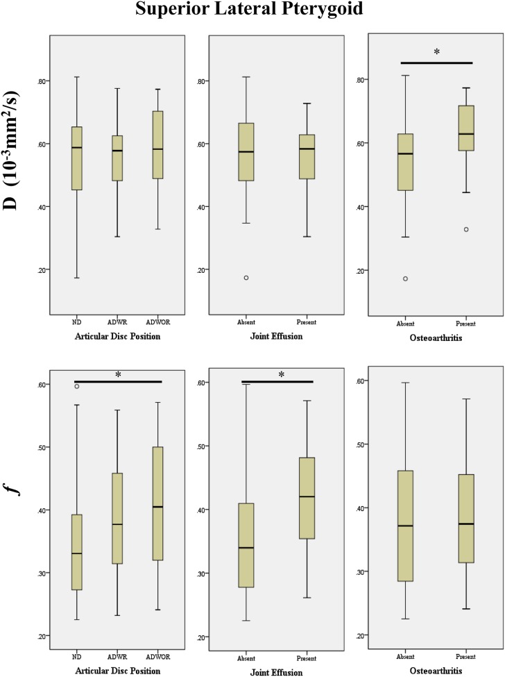
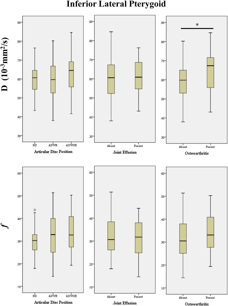
Results: For SLP, the f parameter in TMJs with anterior disc displacement without reduction was significantly higher than that in normal ones (p = 0.015). It was also significantly higher in TMJs with joint effusion than in those without (p = 0.016). On the other hand, for both SLP and ILP, the D parameter significantly increased in TMJs with osteoarthritis compared with those without (p = 0.015 and p = 0.022, respectively).
Conclusions: Pathological changes of LPM in patients with TMD may be quantitatively evaluated by IVIM parameters.
Keywords: diagnostic imaging; diffusion-weighted MRI; masticatory muscles; pterygoid muscle; temporomandibular joint disorders.
Figures
Similar articles
-
A possible etiology of the internal derangement of the temporomandibular joint based on the MRI observations of the lateral pterygoid muscle.Surg Radiol Anat. 2005 Mar;27(1):19-24. doi: 10.1007/s00276-004-0267-6. Epub 2004 Nov 26. Surg Radiol Anat. 2005. PMID: 15750717
-
MR abnormalities of the lateral pterygoid muscle in patients with nonreducing disk displacement of the TMJ.Cranio. 2002 Jul;20(3):209-21. doi: 10.1080/08869634.2002.11746213. Cranio. 2002. PMID: 12150268
-
[Evaluation of Lateral Pterygoid Muscle Contraction in Patients with Temporomandibular Disorders Based on 3D-T2 Weighted Imaging].Zhongguo Yi Xue Ke Xue Yuan Xue Bao. 2021 Aug;43(4):579-583. doi: 10.3881/j.issn.1000-503X.13076. Zhongguo Yi Xue Ke Xue Yuan Xue Bao. 2021. PMID: 34494529 Chinese.
-
Acute dental malocclusion associated with lateral pterygoid muscle partial tear: Case Report and literature review.Cranio. 2024 Jan;42(1):33-39. doi: 10.1080/08869634.2021.1916301. Epub 2021 Apr 17. Cranio. 2024. PMID: 33870872 Review.
-
Imaging features of chondrosarcoma of the temporomandibular joint: report of nine cases and literature review.Clin Radiol. 2020 Nov;75(11):878.e1-878.e12. doi: 10.1016/j.crad.2020.07.016. Epub 2020 Aug 22. Clin Radiol. 2020. PMID: 32843140 Review.
Cited by
-
Relation between Condyle Horizontal Angle and Intercondylar Angle with Disc Displacement in Patients with Temporomandibular Joint Disorders: An MRI Evaluation.Radiol Res Pract. 2023 Sep 8;2023:3846525. doi: 10.1155/2023/3846525. eCollection 2023. Radiol Res Pract. 2023. PMID: 37719870 Free PMC article.
-
Diffusion Tensor Imaging of the Lateral Pterygoid Muscle in Patients with Temporomandibular Joint Disorders and Healthy Volunteers.Korean J Radiol. 2022 Feb;23(2):218-225. doi: 10.3348/kjr.2021.0132. Epub 2021 Oct 1. Korean J Radiol. 2022. PMID: 34668354 Free PMC article.
-
Intravoxel Incoherent Motion Magnetic Resonance Imaging in Skeletal Muscle: Review and Future Directions.J Magn Reson Imaging. 2022 Apr;55(4):988-1012. doi: 10.1002/jmri.27875. Epub 2021 Aug 14. J Magn Reson Imaging. 2022. PMID: 34390617 Free PMC article. Review.
-
Evaluation of lateral pterygoid muscle in patients with temporomandibular joint anterior disk displacement using T1-weighted Dixon sequence: a retrospective study.BMC Musculoskelet Disord. 2022 Feb 8;23(1):125. doi: 10.1186/s12891-022-05079-1. BMC Musculoskelet Disord. 2022. PMID: 35135518 Free PMC article.
-
Temporomandibular Disk Dislocation Impacts the Stomatognathic System: Comparative Study Based on Biexponential Quantitative T2 Maps.J Clin Med. 2022 Mar 15;11(6):1621. doi: 10.3390/jcm11061621. J Clin Med. 2022. PMID: 35329946 Free PMC article.
References
-
- Okeson JP. Management of temporomandibular disorders and occlusion. 7th edn. St. Louis, MO: Mosby; 1998. p. 504.
-
- Sakaguchi-Kuma T, Hayashi N, Fujishiro H, Yamaguchi K, Shimazaki K, Ono T, et al. . An anatomic study of the attachments on the condylar process of the mandible: muscle bundles from the temporalis. Surg Radiol Anat 2016; 38: 461–7. doi: https://doi.org/10.1007/s00276-015-1587-4 - DOI - PubMed
-
- Wilkinson TM. The relationship between the disk and the lateral pterygoid muscle in the human temporomandibular joint. J Prosthet Dent 1988; 60: 715–24. doi: https://doi.org/10.1016/0022-3913(88)90406-4 - DOI - PubMed
-
- Mahan PE, Wilkinson TM, Gibbs CH, Mauderli A, Brannon LS. Superior and inferior bellies of the lateral pterygoid muscle EMG activity at basic jaw positions. J Prosthet Dent 1983; 50: 710–18. doi: https://doi.org/10.1016/0022-3913(83)90214-7 - DOI - PubMed
-
- Wang MQ, Yan CY, Yuan YP. Is the superior belly of the lateral pterygoid primarily a stabilizer? An EMG study. J Oral Rehabil 2001; 28: 507–10. doi: https://doi.org/10.1046/j.1365-2842.2001.00703.x - DOI - PubMed
MeSH terms
LinkOut - more resources
Full Text Sources
Other Literature Sources
Medical
