

Cerebral Cortex Regions Selectively Vulnerable to Radiation Dose-Dependent Atrophy
- PMID: 28333012
- PMCID: PMC5403140
- DOI: 10.1016/j.ijrobp.2017.01.005
Cerebral Cortex Regions Selectively Vulnerable to Radiation Dose-Dependent Atrophy
Abstract
Purpose and objectives: Neurologic deficits after brain radiation therapy (RT) typically involve decline in higher-order cognitive functions such as attention and memory rather than sensory defects or paralysis. We sought to determine whether areas of the cortex critical to cognition are selectively vulnerable to radiation dose-dependent atrophy.
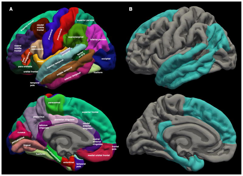
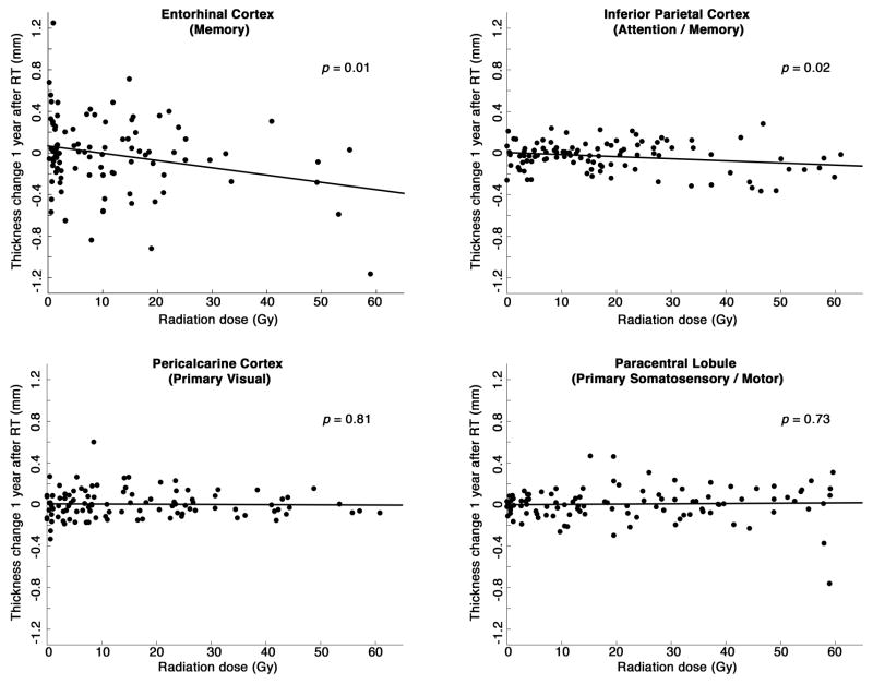
Methods and materials: We measured change in cortical thickness in 54 primary brain tumor patients who underwent fractionated, partial brain RT. The study patients underwent high-resolution, volumetric magnetic resonance imaging (T1-weighted; T2 fluid-attenuated inversion recovery, FLAIR) before RT and 1 year afterward. Semiautomated software was used to segment anatomic regions of the cerebral cortex for each patient. Cortical thickness was measured for each region before RT and 1 year afterward. Two higher-order cortical regions of interest (ROIs) were tested for association between radiation dose and cortical thinning: entorhinal (memory) and inferior parietal (attention/memory). For comparison, 2 primary cortex ROIs were also tested: pericalcarine (vision) and paracentral lobule (somatosensory/motor). Linear mixed-effects analyses were used to test all other cortical regions for significant radiation dose-dependent thickness change. Statistical significance was set at α = 0.05 using 2-tailed tests.
Results: Cortical atrophy was significantly associated with radiation dose in the entorhinal (P=.01) and inferior parietal ROIs (P=.02). By contrast, no significant radiation dose-dependent effect was found in the primary cortex ROIs (pericalcarine and paracentral lobule). In the whole-cortex analysis, 9 regions showed significant radiation dose-dependent atrophy, including areas responsible for memory, attention, and executive function (P≤.002).
Conclusions: Areas of cerebral cortex important for higher-order cognition may be most vulnerable to radiation-related atrophy. This is consistent with clinical observations that brain radiation patients experience deficits in domains of memory, executive function, and attention. Correlations of regional cortical atrophy with domain-specific cognitive functioning in prospective trials are warranted.
Copyright © 2017 Elsevier Inc. All rights reserved.
Conflict of interest statement
This study is not sponsored by industry. No authors have direct conflicts of interest for the submitted work; commercial relationships outside the scope of this work are as follows. Tyler Seibert and Jona Hattangadi-Gluth have received grant funding from Varian Medical Systems for other work. Dr. Seibert has received honoraria from WebMD, Inc. for providing educational content. Vitali Moiseenko reports prior honorarium and travel fees from Varian Medical Systems for a talk outside the submitted work. James Brewer reports stock options and advisory board membership for Human Longevity, Inc. and CorTechs Labs, Inc. Dr. Brewer has also received research grant funding from Navidea Biopharmaceuticals, Inc. and has served on scientific advisory boards for Elan, Bristol-Meyers Squibb, Avanir, Novartis, Genentech, and Eli Lilly. Anders Dale reports grants and non-financial support from General Electric Healthcare (GEHC); equity interest in CorTechs Labs, Inc.; service on the scientific advisory boards for CorTechs Labs, Inc. and Human Longevity, Inc. In addition, Dr. Dale has two patents (US20120280686 and US8160319) licensed to GEHC. The other authors have no conflicts of interest related to this work.
Figures
References
-
- Meyers CA, Brown PD. Role and relevance of neurocognitive assessment in clinical trials of patients with CNS tumors. J Clin Oncol Off J Am Soc Clin Oncol. 2006;24:1305–1309. - PubMed
-
- McDuff SGR, Taich ZJ, Lawson JD, et al. Neurocognitive assessment following whole brain radiation therapy and radiosurgery for patients with cerebral metastases. J Neurol Neurosurg Psychiatry. 2013;84:1384–1391. - PubMed
-
- Saad S, Wang TJC. Neurocognitive Deficits After Radiation Therapy for Brain Malignancies. Am J Clin Oncol. 2015;38:634–640. - PubMed
Publication types
MeSH terms
Grants and funding
LinkOut - more resources
Full Text Sources
Other Literature Sources
Medical
