Observational Study
. 2017 Aug;49(6):555-562.
doi: 10.1002/lsm.22655. Epub 2017 Mar 23.

In vivo multiphoton-microscopy of picosecond-laser-induced optical breakdown in human skin

Affiliations
Observational Study

In vivo multiphoton-microscopy of picosecond-laser-induced optical breakdown in human skin

Mihaela Balu et al. Lasers Surg Med. 2017 Aug.

Abstract

Importance: Improvements in skin appearance resulting from treatment with fractionated picosecond-lasers have been noted, but optimizing the treatment efficacy depends on a thorough understanding of the specific skin response. The development of non-invasive laser imaging techniques in conjunction with laser therapy can potentially provide feedback for guidance and optimizing clinical outcome.

Objective: The purpose of this study was to demonstrate the capability of multiphoton microscopy (MPM), a high-resolution, label-free imaging technique, to characterize in vivo the skin response to a fractionated non-ablative picosecond-laser treatment.

Design, setting, and participants: Two areas on the arm of a volunteer were treated with a fractionated picosecond laser at the Dermatology Clinic, UC Irvine. The skin response to treatment was imaged in vivo with a clinical MPM-based tomograph at 3 hours and 24 hours after treatment and seven additional time points over a 4-week period.

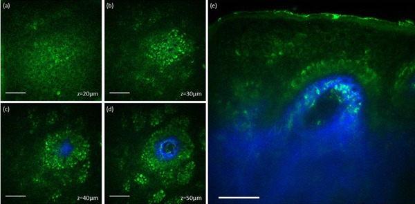
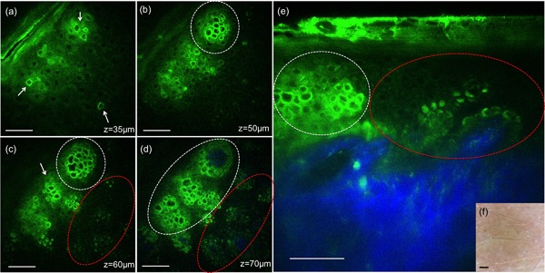
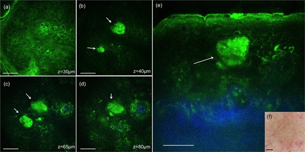
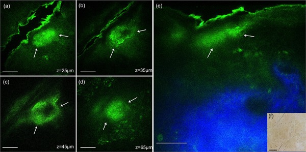
Main outcomes and measures: MPM revealed micro-injuries present in the epidermis. Pigmented cells were particularly damaged in the process, suggesting that melanin is likely the main absorber for laser induced optical breakdown.

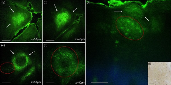
Results: Damaged individual cells were distinguished as early as 3 hours post pico-laser treatment with the 532 nm wavelength, and 24 hours post-treatment with both 532 and 1064 nm wavelengths. At later time points, clusters of cellular necrotic debris were imaged across the treated epidermis. After 24 hours of treatment, inflammatory cells were imaged in the proximity of epidermal micro-injuries. The epidermal injuries were exfoliated over a 4-week period.

Conclusions and relevance: This observational and descriptive pilot study demonstrates that in vivo MPM imaging can be used non-invasively to provide label-free contrast for describing changes in human skin following a fractionated non-ablative laser treatment. The results presented in this study represent the groundwork for future longitudinal investigations on an expanded number of subjects to understand the response to treatment in different skin types with different laser parameters, critical factors in optimizing treatment outcome. Lasers Surg. Med. 49:555-562, 2017. © 2017 Wiley Periodicals, Inc.

Keywords: in vivo imaging; laser induced optical breakdown; noninvasive multiphoton microscopy.


Figures

Figure 1
Figure 1
In vivo MPM images of normal human skin. (a–d) En‐face MPM images (XY scans) showing keratinocytes (green fluorescence) and normal pigmented cells (bright green fluorescence) in the epidermis at z = 20 µm (a), z = 30 µm (b), z = 40 µm (c), and z = 50 µm (d). (e) Cross‐sectional view (XZ scan) representing a vertical plane through the same interrogating volume corresponding to the en‐face images on the left. The image shows normal pigmented cells and collagen (blue). Scale bar is 40 µm in all MPM images.
Figure 2
Figure 2
In vivo MPM images of human skin 3 hours post‐treatment, 532 nm. (a–d) En‐face MPM images (XY scans) showing keratinocytes in the epidermis and individual damaged cells (white arrows) at z = 35 µm (a), clusters of individual damaged cells (white contours) at z = 50 µm (b), 60 µm (c), and 70 µm (d). Normal pigmented cells are outlined by red contours (c and d). (e) Cross‐sectional view (XZ scan) representing a vertical plane through the same interrogating volume corresponding to the en‐face images on the left. The image shows damaged individual cells (white contour) and normal pigmented cells (red contour). The inset shows a close‐up of damaged cells (bright green) adjacent to normal cells (dark green). Scale bar is 40 µm in all MPM images. (f) Clinical image (DermLite FOTO, DermLite Inc.). Scale bar is 1 mm.
Figure 3
Figure 3
In vivo MPM images of human skin 24 hours post‐treatment, 532 nm. (a–d) En‐face MPM images (XY scans) showing keratinocytes in the epidermis, clusters of individual damaged cells (white arrows) at z = 25 µm (a), 35 µm (b), 50 µm (c), and likely inflammatory cells (red contour) at z = 105 µm (d). (e) Cross‐sectional view (XZ scan) representing a vertical plane through the same interrogating volume corresponding to the en‐face images on the left. The image shows a cluster of damaged individual cells (white arrows) and likely inflammatory cells in their proximity (red contour). Scale bar is 40 µm in all MPM images. (f) Clinical image (DermLite FOTO, Dermlite Inc.). Scale bar is 1 mm.
Figure 4
Figure 4
In vivo MPM images of human skin 24 hours post‐treatment, 1064 nm. (a–d) En‐face MPM images (XY scans) showing keratinocytes in the epidermis and clusters of individual damaged cells (white arrows) at z = 30 µm (a), 40 µm (b), 65 µm (c), and z = 80 µm (d). (e) Cross‐sectional view (XZ scan) representing a vertical plane through the same interrogating volume corresponding to the en‐face images on the left. The image shows a cluster of damaged cells (white arrow) in the epidermis. Scale bar is 40 µm in all MPM images. (f) Clinical image (DermLite FOTO, Dermlite Inc.). Scale bar is 1 mm.
Figure 5
Figure 5
In vivo MPM images of human skin 7 days post‐treatment, 532 nm. (a–d) En‐face MPM images (XY scans) showing clusters of cellular necrotic debris in the epidermis (white arrows) at z = 30 µm (a), 40 µm (b) and 65 µm (c), and inflammatory cells at z = 80 µm (d). (e) Cross‐sectional view (XZ scan) representing a vertical plane through the same interrogating volume corresponding to the en‐face images on the left. The image shows a cluster of cellular necrotic debris (white arrows) in the epidermis and likely inflammatory cells (red contour). Scale bar is 40 µm in all MPM images. (f) Clinical image (DermLite FOTO, Dermlite Inc.). Scale bar is 1 mm.
Figure 6
Figure 6
In vivo MPM images of human skin 7 days post‐treatment, 1064 nm. (a–d) En‐face MPM images (XY scans) showing clusters of cellular necrotic debris in the epidermis (white arrows) at z = 25 µm (a), 35 µm (b), 45 µm (c), 65 µm (d). (e) Cross‐sectional view (XZ scan) representing a vertical plane through the same interrogating volume corresponding to the en‐face images on the left. The image shows a cluster of cellular necrotic debris (white arrows) in the epidermis beneath the stratum corneum and normal collagen distribution (blue). Scale bar is 40 µm in all MPM images. (f) Clinical image (DermLite FOTO, Dermlite Inc.). Scale bar is 1 mm.

References

    1. Tanghetti EA. The histology of skin treated with a picosecond alexandrite laser and a fractional lens array. Lasers Surg Med 2016; 48(7):646–652. - PubMed
    1. Habbema L, Verhagen R, Van Hal R, Liu Y, Varghese B. Minimally invasive non‐thermal laser technology using laser‐induced optical breakdown for skin rejuvenation. J Biophotonics 2012; 5(2):194–199. - PMC - PubMed
    1. Habbema L, Verhagen R, Van Hal R, Liu Y, Varghese B. Efficacy of minimally invasive nonthermal laser‐induced optical breakdown technology for skin rejuvenation. Lasers Med Sci 2013; 28(3):935–940. - PMC - PubMed
    1. Balu M, Kelly KM, Zachary CB, et al. Distinguishing between benign and malignant melanocytic nevi by in vivo multiphoton microscopy. Cancer Res 2014; 74(10):2688–2697. - PMC - PubMed
    1. Balu M, Zachary CB, Harris RM, et al. In vivo multiphoton microscopy of basal cell carcinoma. JAMA dermatology 2015; 151(10):1068–1074. - PMC - PubMed

Publication types