Review
. 2017 Mar 16;7(2):485-581.
doi: 10.1002/cphy.c160011.

Smooth Muscle Ion Channels and Regulation of Vascular Tone in Resistance Arteries and Arterioles

Affiliations
Review

Smooth Muscle Ion Channels and Regulation of Vascular Tone in Resistance Arteries and Arterioles

Nathan R Tykocki et al. Compr Physiol. .

Abstract

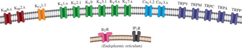
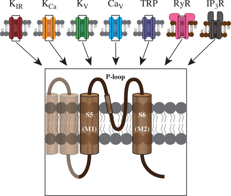
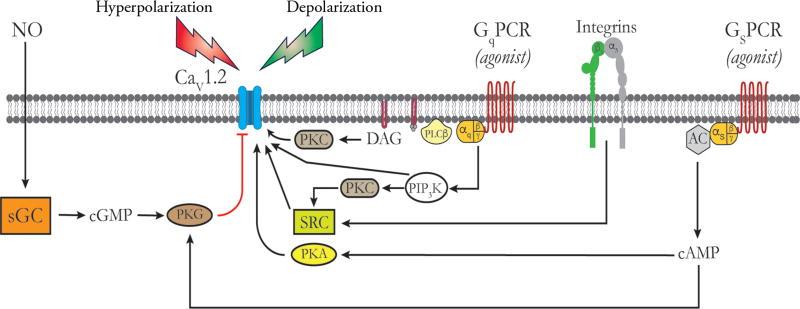
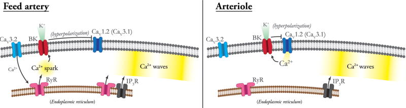
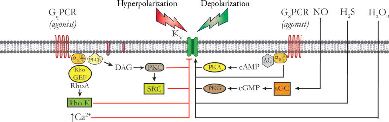
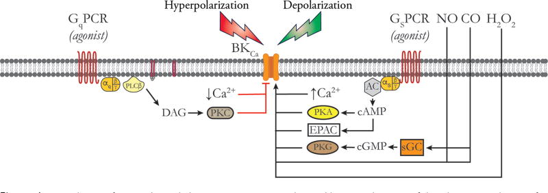
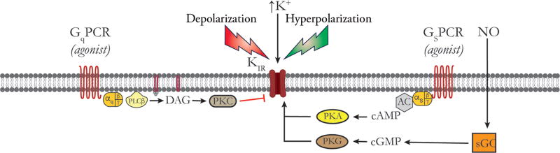
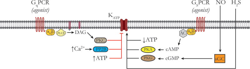
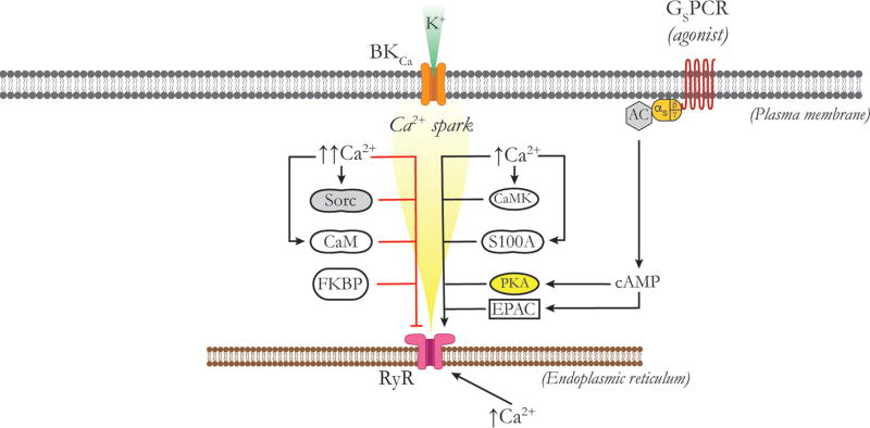
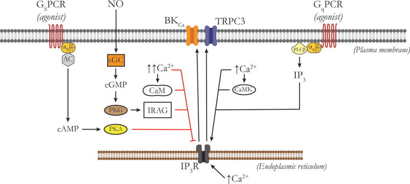
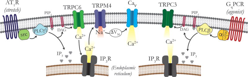
Vascular tone of resistance arteries and arterioles determines peripheral vascular resistance, contributing to the regulation of blood pressure and blood flow to, and within the body's tissues and organs. Ion channels in the plasma membrane and endoplasmic reticulum of vascular smooth muscle cells (SMCs) in these blood vessels importantly contribute to the regulation of intracellular Ca2+ concentration, the primary determinant of SMC contractile activity and vascular tone. Ion channels provide the main source of activator Ca2+ that determines vascular tone, and strongly contribute to setting and regulating membrane potential, which, in turn, regulates the open-state-probability of voltage gated Ca2+ channels (VGCCs), the primary source of Ca2+ in resistance artery and arteriolar SMCs. Ion channel function is also modulated by vasoconstrictors and vasodilators, contributing to all aspects of the regulation of vascular tone. This review will focus on the physiology of VGCCs, voltage-gated K+ (KV) channels, large-conductance Ca2+-activated K+ (BKCa) channels, strong-inward-rectifier K+ (KIR) channels, ATP-sensitive K+ (KATP) channels, ryanodine receptors (RyRs), inositol 1,4,5-trisphosphate receptors (IP3Rs), and a variety of transient receptor potential (TRP) channels that contribute to pressure-induced myogenic tone in resistance arteries and arterioles, the modulation of the function of these ion channels by vasoconstrictors and vasodilators, their role in the functional regulation of tissue blood flow and their dysfunction in diseases such as hypertension, obesity, and diabetes. © 2017 American Physiological Society. Compr Physiol 7:485-581, 2017.


Figures

Figure 1
Figure 1
Principal ion channels expressed in vascular SMCs. In the plasma membrane (gray), the following channels are expressed: at least two members of the inward-rectifier K+ channel (KIR) family; large-conductance, Ca2+-activated K+ channels (KCa 1.1); at least six members of the voltage-dependent K+ channel (Kv) family; at least two voltage-dependent Ca2+ channels (Cav); and a number of TRP channels. In the endoplasmic reticular membrane (brown), RyRs and IP3R are expressed.
Figure 2
Figure 2
Pore-forming subunits of ion channels. All ion channels share a similar topology, wherein the S5 and S6 transmembrane domains (M1 and M2 for KIR channels) form the ion-permeable pore. These two domains are linked by a pore-loop (P-loop), which contains multiple residues responsible for regulating pore function and ion selectivity. See text for details.
Figure 3
Figure 3
Regulation of CaV 1.2 channels by vasoconstrictors and vasodilators. Schematic of the plasma membrane of a vascular SMC showing, from left to right, a CaV 1.2 channel, a Gq-protein coupled receptor (GqPCR), an α5β1 Integrin and a Gs-protein coupled receptor (GSPCR). Black lines and arrows indicate stimulation, activativation or increases; red lines indicate inhibition. Pathways to the right of the CaV 1.2 activate these channels, while those to the left are inhibitory. Membrane depolarization due to opening of membrane channels that conduct Na+, Ca2+, or Cl or due to closue of K+ channels represents the major stimulus for opening CaV 1.2 channels. Vasoconstictor agonists that act through GqPCRs norepinephrine, endothelin, angiotensin II, 5-HT, etc.) are coupled to phospholipase Cβ (PLCβ), which acts on q membrane phophoinositol bisphosphate to form diacylglycerol (DAG), which, in the presence of Ca2+, activates PKC. PKC phosphorylates CaV 1.2 to increase its open-state probability. GqPCR activation can also stimulate phosphatidyl inostitol trisphosphate kinase (PIP3K), which acts on novel PKCs to activate the tyrosine kinase SRC, as shown. SRC phosphorylates CaV 1.2 channels, increasing their activity. Activation of CaV 1.2 by PIP3K independent of PKC and SRC has also been reported. SRC can also be activated by activated inetgrins as shown, also increasing CaV 1.2 activity. Agonists for GsPCR (isoproterenol, adenosine, prostacyclin, CGRP, etc.) activate adenylate cyclase (AC) to increase the formation of cAMP which activates PKA. PKA phosphorylates CaV 1.2 to increase the activity of this channel. Membrane hyperpolarization due to opening of K+ channels or closure of channels conducting Na+, Ca2+, or Cl represents the main stimulus for deactivation of CaV 1.2 channels. In addition, nitric oxide (NO) acting through soluble guanylate cyclase (sGC), and other agents that increase cGMP, activate protein kinase G (PKG) which can phosphorylate CaV 1.2 channels to decrease their activity. In addition, high levels of cAMP can transactivate PKG accounting for the inhibitory effects of high levels of activation of GsPCR or direct activators of AC such as forskolin on CaV 1.2 channel activity. See text for details and references.
Figure 4
Figure 4
Calcium signaling in feed arteries versus downstream arterioles. Feed arteries display both Ca2+ sparks and Ca2+ waves, as shown. Ca2+ sparks in feed arteries arise from RyRs that may be activated by Ca2+ influx through CaV 3.2 channels via Ca2+-induced Ca2+ release. In feed arteries, Ca2+ sparks activate BKCa channels, hyperpolarizing the membrane and deactivating CaV 1.2 channels, which contributes to the negative feedback regulation of myogenic tone. Ca2+ waves in feed arteries depend on the activity of both RyRs and IP3Rs. In arterioles, Ca2+ influx through CaV 1.2 and other VGCCs provides the Ca2+ signal for activation of BKCa channels and the negative feedback regulation of membrane potential and VGCC activity. Ca2+ waves in arterioles depend solely on the activity of IP3R. RyRs are expressed in arteriolar SMCs but are silent under resting conditions. See text for details.
Figure 5
Figure 5
Regulation of KV channels by vasoconstrictors and vasodilators. Schematic of the plasma membrane of a vascular SMC showing, from left to right, a Gq-protein-coupled receptor (GqPCR), associated G-proteins and phospholipase C-β (PLCβ); a generic KV channel; and a Gs-protein-coupled receptor, associated G-proteins and adenylate cyclase (AC). Black lines and arrows indicate stimulation, activation or increases; red lines indicate inhibition. Pathways to the right of the KV channel activate these channels, while those to the left are inhibitory. Membrane depolarization due to opening of membrane channels that conduct Na+, Ca2+, or Cl or due to closure of other K+ channels represents the major stimulus for opening KV channels. Vasodilator agonists that act at GsPCR (isoproterenol, adenosine, prostacyclin, CGRP, etc.), stimulate the formation of cAMP, activation of PKA and phosphorylation of KV channels leading to their activation. In addition, the Gβγ-subunits can directly interact with some KV channels also leading to their activation. NO, acting through sGC, and other vasodilators that stimulate the production of cGMP, activeate PKG, phosphorylating KV channels and increasing their activity. Other vasodilators, such as H2S and H2O2 also can activate KV channels as shown. Hyperpolarization, induced by opening of other K+ channels or closure of channels conducting Na+, Ca2+, or Cl, represents the major stimulus for closure of KV channels. In addition, vasoconstrictors that act through Gq-coupled receptors can inhibit KV channels through several mecahisms including: (A) the activation of PLCβ, the formation of DAG and activation of PKC; (B) PKC-dependent activation of the tyrosine kinase SRC; (C) Rho-guanine-nuclotide exchange factor (Rho-GEF)-dependent activation of RhoA and Rho kinase (Rho K); and (D) agonist-induced increases in intracellular Ca2+. See text for more information.
Figure 6
Figure 6
Regulation of BKCa channels by vasoconstrictors and vasodilators. Schematic of the plasma membrane of a vascular SMC showing, from left to right, a Gq-protein-coupled receptor (GqPCR), associated G-proteins and PLCβ; a BKCa channel; and a Gs-protein-coupled receptor, associated G-proteins and AC. Black lines and arrows indicate stimulation, activation or increases; red lines indicate inhibition. Pathways to the right of the BKCa channel activate these channels, while those to the left are inhibitory. Membrane depolarization due to opening of membrane channels that conduct Na+, Ca2+, or Cl or due to closure of other K+ channels as well as increases in subsarcolemmal Ca2+ are the major stimulae for opening BKCa channels. Vasodilator agonists that act at GsPCR (isoproterenol, adenosine, prostacyclin, CGRP, etc.), stimulate the formation of cAMP, activation of PKA and phosphorylation of BKCa channels leading to their activation. Vasodilators that lead to increased production of cAMP als may active BKCa channels through exchange EPACs. NO, acting through sGC, and other vasodilators that stimulate the production of cGMP, activate PKG, phosphorylating BKCa channels and increasing their activity. Other vasodilators, such as H2S and H2O2 also can activate KV channels as shown. NO or carbon monoxide (CO) also may directly interact with BKCa channels or associated heme-proteins to increase channel activity. BKCa channels also are activated by H2O2. Conversely, hyperpolarization, induced by opening of other K+ channels or closure of channels conducting Na+, Ca2+, or Cl, and/or a fall in subsarcolemmal Ca2+ represent the major stimulae for closure of KV channels. In addition, vasoconstrictors that act through Gq-coupled receptors can inhibit BKCa channels through activation of PLCβ, the formation of diacylglycerol (DAG) and activation of PKC. See text for more information.
Figure 7
Figure 7
Regulation of KIR channels by vasoconstrictors and vasodilators. Schematic of the plasma membrane of a vascular SMC showing, from left to right, a Gq-protein-coupled receptor (GqPCR), associated G-proteins and PLCβ; a KIR channel; and a Gs-protein-coupled receptor, associated G-proteins and AC. Black lines and arrows indicate stimulation, activation or increases; red lines indicate inhibition. Hyperpolarization induced by the activation of other K+ channels, or the closure of channels conducting Na+, Ca2+, or Cl and/or increases in extracellular K+ concentration are the major stimuli for activation of vascular SMC KIR channels. In addition, vasodilators that act at GsPCRs (isoproterenol, adenosine, prostacyclin, CGRP, etc.), stimulate AC, increase the production of cAMP and activate PKA lead to activation of KIR channels. Similarly, NO, acting through sGC to increase production of cGMP, activated protein kinase G which can activate KIR channels. Conversely, membrane depolarization due to closure of other K+ channels or opening of channels that conduct Na+, Ca2+, or Cl will close KIR channels. Vasoconstrictors that act through GqPCRs (norepinephrine, endothelin, angiotensin II, 5-HT, etc.) to activate PLCβ, the production of DAG and PKC activation lead to closure of KIR channels. See text for more information.
Figure 8
Figure 8
Regulation of KATP channels by vasoconstrictors and vasodilators. Schematic of the plasma membrane of a vascular SMC showing, from left to right, a Gq-protein-coupled receptor (GqPCR), associated G-proteins and PLCβ; a KATP channel; and a Gs-protein-coupled receptor, associated G-proteins and AC. Black lines and arrows indicate stimulation, activation or increases; red lines indicate inhibition. These channels can be activated by a fall in intracellular ATP in the environment of these channels. In addition, vasodilators that act at GSPCRs (isoproterenol, adenosine, prostacyclin, CGRP, etc.), stimulate AC, increase the production of cAMP and activate PKA lead to activation of KATP channels. Similarly, NO, acting through sGC to increase production of cGMP, activating PKG which can activate KATP channels. These channels also can be activated by H2S, as shown. Conversely, increases in ATP close KATP channels. Vasoconstrictors that act through GqPCRs (norepinephrine, endothelin, angiotensin II, serotonin, etc.) to activate PLCβ, the production of DAG and PKC activation will lead to closure of KIR channels. Increases in intracellular Ca2+ that accompany SMC stimulation by vasoconstrictors activates protein phosphatase 2B (calcineurin), which also closes KATP channels by dephosphorylation. See text for more information.
Figure 9
Figure 9
Regulation of RyRs. Schematic of the plasma membrane and the ER membrane of a vascular SMC showing, from left to right in the plasma membrane, a BKCa channel and a Gs-protein-coupled receptor, associated G-proteins and AC, and a RyR in the membrane of the ER. Moderate increases in cytoplasmic Ca2+ in the environment of a RyR, or increases in the concentration of Ca2+ in the lumen of the ER are the primary stimulae for activation of RyRs. Activation of RyR by cytosolic Ca2+ is mediated by direct actions of Ca2+ on the channels, through activation of calcium-calmodulin-dependent protein kinase (CaMK) and phosphorylation of the channels, or interactions of Ca2+ with the Ca2+-binding protein S100A which competes with calmodulin for binding to the RyR. Vasodilators that act at GsPCRs (isoproterenol, adenosine, prostacyclin, CGRP, etc.), activate AC to increase production of cAMP which then can activate PKA to phosphorylate RyRs and increase their activity increasing the frequency of production of Ca2+ sparks. Elevated cAMP can also increase RyR activity through activation of EPACs. The increase in RyR-depedent Ca2+ spark activity is transduced into membrane hyperpolarization and vasodilation through activation of overlying BKCa channels, as shown. Conversely, high levels of intracellular Ca2+ inhibit RyR activity through mechanisms involving the Ca2+-binding proteins sorcin (SORC) or calmodulin (CaM). The activity of RyRs also may be decreased by interactions with FK-506 binding proteins 12 and 12A FKBPs. See text for more information.
Figure 10
Figure 10
Regulation of IP3 receptors by vasoconstrictors and vasodilators. Schematic of the plasma membrane and the ER membrane of a vascular SMC showing, from left to right in the plasma membrane, a Gs-protein-coupled receptor, associated G-proteins and AC, a BKCa channel, a TRPC3 channel and a Gq-protein-coupled receptor (GqPCR), associated G-proteins and phospholipase C-β (PLCβ), and an IP3 receptor (IP3R) in the membrane of the ER. Increases in IP3 and moderate increases in cytoplasmic Ca2+ in the environment of an IP3R are the primary stimulae for activation. Activation of IP3Rs by cytosolic Ca2+ is mediated by direct actions of Ca2+ on the channels, or through activation of calcium-calmodulin-dependent protein kinase (CaMK) and phosphorylation of the channels. Vasoconstrictors acting through GqPCRs and activation of PLCβ increase the production of IP3, stimulating Ca2+ release through IP3R. Activated IP3Rs have been shown to physically interact with, and activate plasma membrane BKCa and TRPC3 channels, as shown. Conversely, high levels of intracellular Ca2+ inhibit IP3R activity through mechanisms involving the Ca2+ binding protein, CaM. NO, through activation of soluble guanylate cyclase, increased production of cGMP and activation of PKG phosphorylates IRAG which inhibits IP3R activity. Vasodilators that act at GsPCRs (isoproterenol, adenosine, prostacyclin, CGRP, etc.), activate AC to increase producting of cAMP which then can activate PKA to phosphorylate IP3Rs to decrease their activity. See text for more information.
Figure 11
Figure 11
TRP channel regulation of myogenic and agonist-induced smooth muscle contractility. Increased intravascular pressure activates a stretch-sensitive Gq-protein-coupled-receptor (GqPCR; e.g., angiotensin type 1 receptor, AT1R), which then causes the hydrolysis of PIP2 by phospholipase C- γ1 (PLCγ1) to form DAG and IP3. DAG activates TRPC6 channels to increase cytosolic Ca2+ concentration, combined with IP3-mediated Ca2+ release through IP3Rs in the ER. This local increase in cytosolic Ca2+ concentration activates TRPM4-dependent Na+ influx, membrane depolarization, and opening of Cav 1.2 channels. This results in SMC contraction (myogenic tone). A similar mechanism is activated in response to a GqPCR agonist, through activation of PLCβ and TRPC3 channels. Figure adapted, with permission, from Earley and Brayden (361). See text for more information.

References

    1. Aalkjaer C, Nilsson H. Vasomotion: Cellular background for the oscillator and for the synchronization of smooth muscle cells. Br J Pharmacol. 2005;144:605–616. - PMC - PubMed
    1. Abbink EJ, Wollersheim H, Netten PM, Russel FG, Lutterman JA, Smits P. Microcirculatory effects of KATP channel blockade by sulphonylurea derivatives in humans. Eur J Clin Invest. 2002;32:163–171. - PubMed
    1. Abd El-Rahman RR, Harraz OF, Brett SE, Anfinogenova Y, Mufti RE, Goldman D, Welsh DG. Identification of L- and T-type Ca2+ channels in rat cerebral arteries: Role in myogenic tone development. Am J Physiol Heart Circ Physiol. 2013;304:H58–H71. - PMC - PubMed
    1. Abdel-Latif AA. Cross talk between cyclic nucleotides and polyphos-phoinositide hydrolysis, protein kinases, and contraction in smooth muscle. Exp Biol Med (Maywood) 2001;226:153–163. - PubMed
    1. Abdi A, Mazzocco C, Legeron FP, Yvert B, Macrez N, Morel JL. TRPP2 modulates ryanodine- and inositol-1,4,5-trisphosphate receptors-dependent Ca2+ signals in opposite ways in cerebral arteries. Cell Calcium. 2015;58:467–475. - PubMed

Publication types

MeSH terms

LinkOut - more resources