. 2017 Apr 21;35(4):237-247.
doi: 10.1080/07357907.2017.1292517. Epub 2017 Mar 23.

Cyclooxygenase-2 Induction by Amino Acid Deprivation Requires p38 Mitogen-Activated Protein Kinase in Human Glioma Cells

Affiliations

Cyclooxygenase-2 Induction by Amino Acid Deprivation Requires p38 Mitogen-Activated Protein Kinase in Human Glioma Cells

Zhiwen Li et al. Cancer Invest. .

Abstract

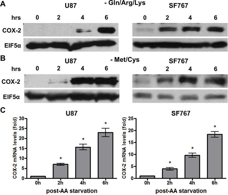
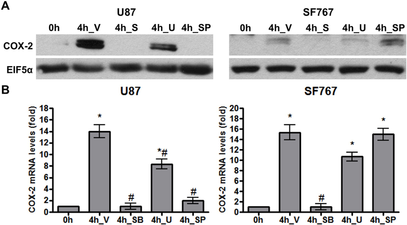
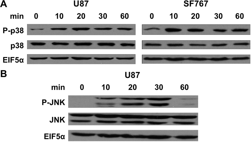
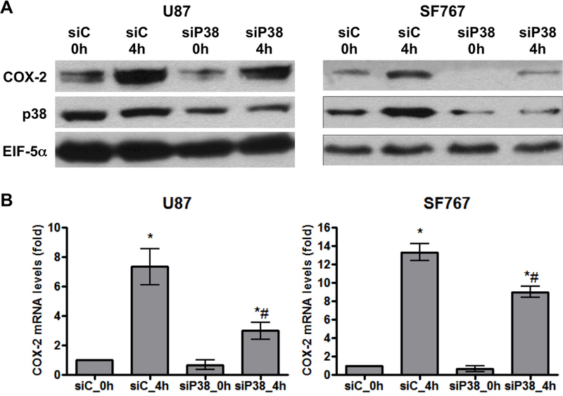
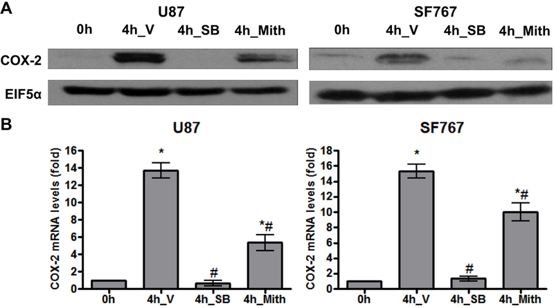
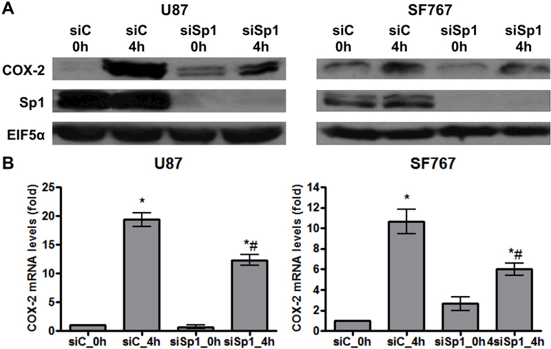
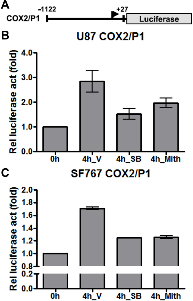
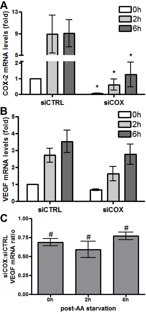
Glioblastomas (GBMs) are malignant brain tumors that can outstrip nutrient supplies due to rapid growth. Cyclooxygenase-2 (COX-2) has been linked to GBMs and may contribute to their aggressive phenotypes. Amino acid starvation results in COX-2 mRNA and protein induction in multiple human glioma cell lines in a process requiring p38 mitogen-activated protein kinase (p38-MAPK) and the Sp1 transcription factor. Increased vascular endothelial growth factor expression results from starvation-dependent COX-2 induction. These data suggest that COX-2 induction with amino acid deprivation may be a part of the adaptation of glioma cells to these conditions, and potentially alter cellular response to anti-neoplastic therapy.

Keywords: COX-2; Glioblastomas; VEGF; amino acid deprivation; p38-MAPK.


Conflict of interest statement

The authors report no conflicts of interest.

Figures

Figure 1.
Figure 1.
AA starvation induces COX-2 expression. Indicated cells were cultured in media lacking (A) glutamine/arginine/lysine (Gln/Arg/Lys) or (B/C) methionine/cysteine (Met/Cys). Cells were subjected to starvation conditions for various times and (A/B) probed for COX-2 protein expression on immunoblots or (C) COX-2 mRNA expression by real-time PCR. EIF5α and cyclophilin A served as normalization controls for the immunoblots and real-time PCR assays, respectively. Blots are representative of three independent experiments. Graphs show average value of three independent experiments with error bar representing ± one SEM. * indicate statistically significant (p < 0.05) difference by student’s t-test compared with initial time point.
Figure 2.
Figure 2.
AA starvation-dependent COX-2 induction is inhibited by p38-MAPK inhibition. Indicated cells were pretreated for one hour with DMSO (V, vehicle), SB202190 (SB, 20 μM, p38-MAPK inhibitor), U0126 (U, 10 μM, MEK inhibitor), or SP600125 (SP, 30 μM, JNK inhibitor) and then AA starved for 4 hours. COX-2 (A) protein and (B) mRNA expression were assessed. EIF5α and cyclophilin A again served as normalization controls. Blots are representative of three independent experiments. Graphs show average value of three independent experiments with error bar representing ± one SEM. * and # indicate statistically significant (p < 0.05) difference by t-test compared with the initial and V-treated 4 hour time points, respectively.
Figure 3.
Figure 3.
Activation of p38-MAPK and JNK by AA starvation. Indicated cells were harvested at the indicated times after AA starvation and assessed for expression of (A) p38-MAPK and phosphorylated p38-MAPK (P-p38) or (B) JNK and phosphorylated JNK (P-JNK) by immunoblot analysis. EIF5α again served as normalization controls. Blots are representative of three independent experiments.
Figure 4.
Figure 4.
Knockdown of p38-MAPK reduces AA starvation-induced COX-2 expression. Indicated cells were subjected to AA starvation for 4 hours two days after transient transfection with siRNA against control (siC) or p38-MAPK (siP38). COX-2 (A) protein and (B) mRNA expression were assessed. EIF5α and cyclophilin A again served as normalization controls. Blots are representative of three independent experiments. Graphs show the average value of three independent experiments with error bar representing ± one SEM. * and # indicate statistically significant (p < 0.05) difference by t-test compared with the corresponding initial and siC-transfected 4 hour time points, respectively.
Figure 5.
Figure 5.
AA starvation-dependent COX-2 induction is reduced with Sp1 inhibition. Indicated cells were pretreated for one hour with V, SB (20 μM), or mithramycin (Mith, 0.5 μM, Sp1 inhibitor) and then AA starved for 4 hours. COX-2 (A) protein and (B) mRNA expression were assessed. EIF5α and cyclophilin A again served as normalization controls. Blots are representative of three independent experiments. Graphs show the average value of three independent experiments with error bar representing ± one SEM. * and # indicate statistically significant (p < 0.05) difference by t-test compared with the initial and V-treated 4 hour time points, respectively.
Figure 6.
Figure 6.
Knockdown of Sp1 reduces AA starvation-induced COX-2 expression. Indicated cells were subjected to AA starvation for 4 hours two days after transient transfection with siRNA against control (siC) or Sp1 (siSp1). COX-2 (A) protein and (B) mRNA expression were assessed. EIF5α and cyclophilin A again served as normalization controls. Blots are representative of three independent experiments. Graphs show the average value of three independent experiments with error bar representing ± one SEM. * and # indicate statistically significant (p < 0.05) difference by t-test compared with the corresponding initial and siC-transfected 4 hour time points, respectively.
Figure 7.
Figure 7.
AA starvation induces COX-2 promoter activity. Indicated cells were subjected to AA starvation for 4 hours two days after transient transfection with the COX2/P1-luciferase reporter plasmid. (A) The COX-2 promoter expression cassette is shown. (B) U87 or (C) SF767 cells were assessed for luciferase expression with values expressed as fold induction relative to the initial time point. Cells were treated with V, SB (20 μM) or Mith (0.5 μM), as indicated. Graphs show the average value of two independent experiments with error bar representing ± one SEM.
Figure 8.
Figure 8.
AA starvation-induced VEGF expression is dependent on COX-2 expression. U87 cells were subjected to AA starvation for the indicated times after transient transfection with siRNA against control (siCTRL) or COX-2 (siCOX). COX-2 and VEGF mRNA levels were determined at each time point by real-time PCR. (A) COX-2 and (B) VEGF mRNA expression at 0, 2 and 6 hours post-starvation (0h, 2h and 6h) in siCTRL and siCOX cells are shown relative to values from siCTRL cells that have not undergone starvation (0h, arbitrarily set at a value of 1). * indicate statistically significant (p < 0.05) differences by paired t-test compared with the corresponding siCTRL time points. (C) Relative reduction in VEGF mRNA expression is shown expressed as a ratio of values from siCOX to siCTRL cells. # indicate statistically significant reduction in VEGF expression by paired one-tailed t-test at each time point. Graphs show the average value of four independent experiments with error bar representing ± one SEM.

Similar articles

Cited by

References

    1. Smith WL; DeWitt DL; Garavito RM Cyclooxygenases: structural, cellular, and molecular biology. Annu Rev Biochem 2000. 69, 145–82. - PubMed
    1. Dandekar DS; Lokeshwar BL Inhibition of cyclooxygenase (COX)-2 expression by Tet-inducible COX-2 antisense cDNA in hormone-refractory prostate cancer significantly slows tumor growth and improves efficacy of chemotherapeutic drugs. Clin Cancer Res 2004. 10, 8037–47. - PubMed
    1. Kishi K; Petersen S; Petersen C; Hunter N; Mason K; Masferrer JL; Tofilon PJ; Milas L Preferential enhancement of tumor radioresponse by a cyclooxygenase-2 inhibitor. Cancer Res 2000. 60, 1326–31. - PubMed
    1. Oshima M; Dinchuk JE; Kargman SL; Oshima H; Hancock B; Kwong E; Trzaskos JM; Evans JF; Taketo MM Suppression of intestinal polyposis in Apc delta716 knockout mice by inhibition of cyclooxygenase 2 (COX-2). Cell 1996. 87, 803–9. - PubMed
    1. Tsujii M; Kawano S; DuBois RN Cyclooxygenase-2 expression in human colon cancer cells increases metastatic potential. Proc Natl Acad Sci U S A 1997. 94, 3336–40. - PMC - PubMed

MeSH terms

LinkOut - more resources