. 2017 Aug;36(8):1676-1685.
doi: 10.1109/TMI.2017.2686006. Epub 2017 Mar 22.

Constrained Inversion and Spectral Unmixing in Multispectral Optoacoustic Tomography

Constrained Inversion and Spectral Unmixing in Multispectral Optoacoustic Tomography

Lu Ding et al. IEEE Trans Med Imaging. 2017 Aug.

Abstract

Accurate extraction of physical and biochemical parameters from optoacoustic images is often impeded due to the use of unrigorous inversion schemes, incomplete tomographic detection coverage, or other experimental factors that cannot be readily accounted for during the image acquisition and reconstruction process. For instance, inaccurate assumptions in the physical forward model may lead to negative optical absorption values in the reconstructed images. Any artifacts present in the single wavelength optoacoustic images can be significantly aggravated when performing a two-step reconstruction consisting in acoustic inversion and spectral unmixing aimed at rendering the distributions of spectrally distinct absorbers. We investigate a number of algorithmic strategies with non-negativity constraints imposed at the different phases of the reconstruction process. Performance is evaluated in cross-sectional multispectral optoacoustic tomography recordings from tissue-mimicking phantoms and in vivo mice embedded with varying concentrations of contrast agents. Additional in vivo validation is subsequently performed with molecular imaging data involving subcutaneous tumors labeled with genetically expressed iRFP proteins and organ perfusion by optical contrast agents. It is shown that constrained reconstruction is essential for reducing the critical image artifacts associated with inaccurate modeling assumptions. Furthermore, imposing the non-negativity constraint directly on the unmixed distribution of the probe of interest was found to maintain the most robust and accurate reconstruction performance in all experiments.


Figures

Fig. 1
Fig. 1
Normalized extinction (absorption) spectra of the different intrinsic tissue chromophores and optical contrast agents considered in this study.
Fig. 2
Fig. 2
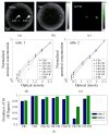
Unmixing results for the phantom with background ink absorption and two insertions (tubes) containing AF750 dye. a) Optoacoustic image acquired at 740 nm with 2.5 OD of AF750 insertion. b) Unmixed image corresponding to the ink component obtained with the CR-CM method. c) Unmixed image corresponding to the AF750 component obtained with the CR-CM method. d)–e) Normalized unmixed concentration (pixel values of the unmixed image) within the tubes as a function of the optical density of AF750. f) The R2 values, representing quality of the linear fit in d) and e).
Fig. 3
Fig. 3
Unmixing results for the in vivo (mouse 1) experiment. a) Unmixed distribution of AF750 obtained with the CR-CM method. b) Unmixed distribution of GNR obtained with the CR-CM method. c)–d) Unmixed optoacoustic signal within the tubes as a function of the optical density of AF750 and GNR, respectively, normalized to the maximum value for the corresponding slices. e)–f) Statistical analysis of the linear fit of the curves in c)–d). All 10 imaged cross-sections were taken into account.
Fig. 4
Fig. 4
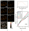
Cross-talk artifacts evaluation for the AF750 probe unmixing in mouse 1. a) Unmixed distribution of AF750 (1.9 OD) for an intestinal region slice using the different reconstruction and unmixing methods. b) Blind spectrum of AF750 used for unmixing. c) Signal to cross-talk ratios as a function of the optical density of AF750 averaged over all 10 imaged cross-sections.
Fig. 5
Fig. 5
Cross-talk artifacts evaluation for the GNR unmixing in mouse 1. a) Unmixed distribution of GNR (1.5 OD) for an intestinal/leg region. b) Blind spectrum of GNR used for unmixing. c) Signal to cross-talk ratios as a function of the optical density of GNR averaged over all 10 imaged cross-sections.
Fig. 6
Fig. 6
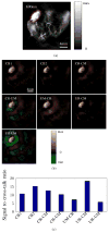
Results of the in vivo iRFP unmixing experiment in mouse 2. a) Single wavelength optoacoustic image (gray scale) acquired at 690 nm. b) Unmixed distributions of iRFP obtained using different methods (brown-green scale). c) Cross-talk performance of different methods - the unmixed iRFP signal is assumed to be confined within the red region marked in a).
Fig. 7
Fig. 7
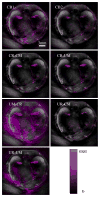
Unmixed distributions of IRDye800CW obtained using the different non-negative constraints. The probe distribution (represented on a purple scale) is superimposed onto the single wavelength optoacoustic images acquired at 850 nm showing accumulation in the renal medulla while clearing through kidneys.

Similar articles

Cited by

References

    1. Razansky D, Distel M, Vinegoni C, Ma R, Perrimon N, Köster RW, Ntziachristos V. Multispectral opto-acoustic tomography of deep-seated fluorescent proteins in vivo. Nature Photonics. 2009;3(7):412–417.
    1. Yao J, Wang LV. Photoacoustic tomography: fundamentals, advances and prospects. Contrast media & molecular imaging. 2011;6(5):332–345. - PMC - PubMed
    1. Jeon M, Kim J, Kim C. Multiplane spectroscopic whole-body photoacoustic imaging of small animals in vivo. Medical & biological engineering & computing. 2016;54(2–3):283–294. - PubMed
    1. Nie L, Chen X. Structural and functional photoacoustic molecular tomography aided by emerging contrast agents. Chemical Society Reviews. 2014;43(20):7132–7170. - PMC - PubMed
    1. Deán-Ben XL, Razansky D. Adding fifth dimension to optoacoustic imaging: volumetric time-resolved spectrally enriched tomography. Light: Science & Applications. 2014;3(1):e137.