. 2017 Mar 23;12(3):e0174006.
doi: 10.1371/journal.pone.0174006. eCollection 2017.

Synthesis and bioevaluation of N,4-diaryl-1,3-thiazole-2-amines as tubulin inhibitors with potent antiproliferative activity

Affiliations

Synthesis and bioevaluation of N,4-diaryl-1,3-thiazole-2-amines as tubulin inhibitors with potent antiproliferative activity

Maolin Sun et al. PLoS One. .

Abstract

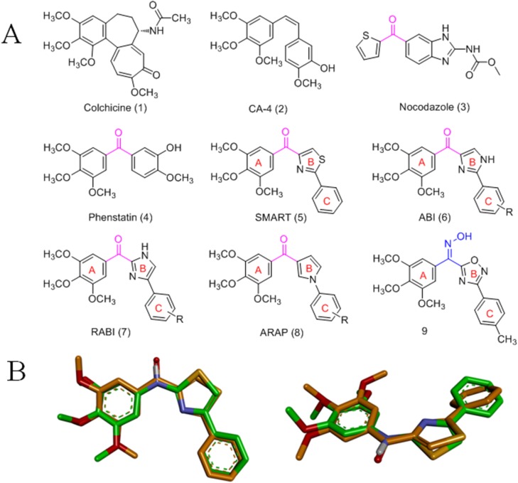
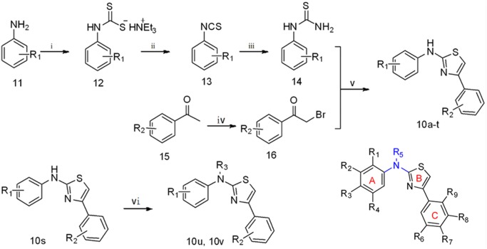
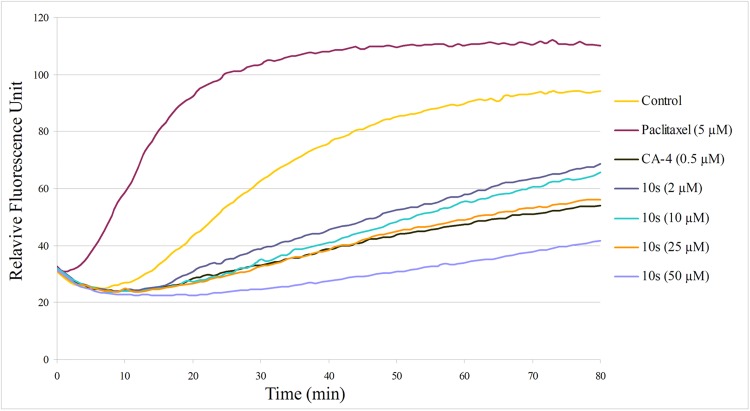
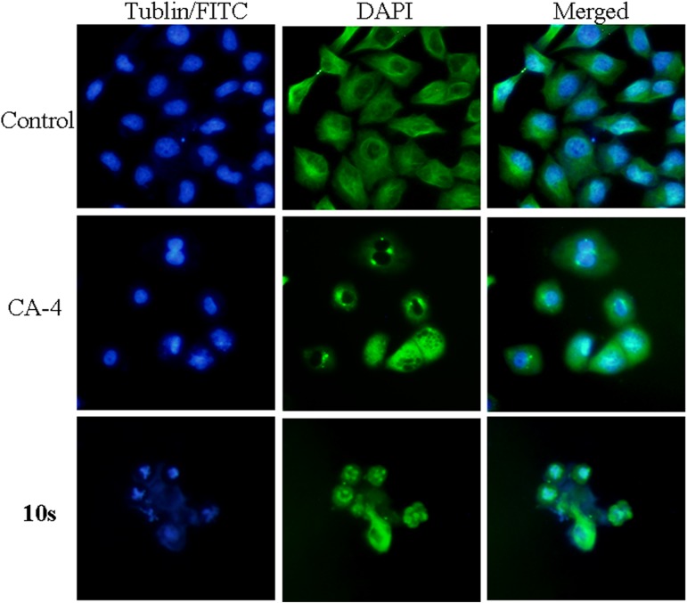
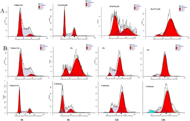
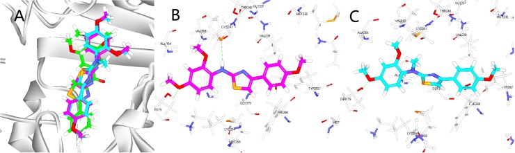
A series of N,4-diaryl-1,3-thiazole-2-amines containing three aromatic rings with an amino linker were designed and synthesized as tubulin inhibitors and evaluated for their antiproliferative activity in three human cancer cell lines. Most of the target compounds displayed moderate antiproliferative activity, and N-(2,4-dimethoxyphenyl)-4-(4-methoxyphenyl)-1,3-thiazol-2-amine (10s) was determined to be the most potent compound. Tubulin polymerization and immunostaining experiments revealed that 10s potently inhibited tubulin polymerization and disrupted tubulin microtubule dynamics in a manner similar to CA-4. Moreover, 10s effectively induced SGC-7901 cell cycle arrest at the G2/M phase in both concentration- and time-dependent manners. The molecular docking results revealed that 10s could bind to the colchicine binding site of tubulin.


Conflict of interest statement

Competing Interests: The authors have declared that no competing interests exist.

Figures

Fig 1
Fig 1
(A) Structures of known tubulin inhibitors. (B) Superposition of the skeletal structure of target compounds 10a-m and SMART. All density functional theory calculations and geometry optimizations were performed using the Gaussian 09 software.
Fig 2
Fig 2
Reagents and conditions. (i) CS2, Et3N, Et2O, 25°C; (ii) I2, Et3N, EtOAc, 0°C; (iii) NH3·H2O; (iv) CuBr2, CHCl3, EtOAc; (v) EtOH, MW 80°C; (vi) 1) CH3I, DMF, 25°C or 2) Ac2O, 50°C.
Fig 3
Fig 3. Effects of 10s on tubulin polymerization.
Tubulin was pre-incubated for 1 min with 10s at 2 μM, 10 μM, 25 μM and 50 μM, CA-4 at 0.5 μM, paclitaxel at 5 μM or vehicle DMSO (control) at room temperature before GTP was added to start the tubulin polymerization reactions. The reaction was monitored at 37°C.
Fig 4
Fig 4. Immunostaining of tubulin assembly in SGC-7901 cells.
SGC-7901 cells were treated with 10s (0.72 μM) for 48 h. Tubulin assembly was stained with FITC (left panel) and DAPI (middle panel). A merge of the two panels is shown on the right. Images were taken using a confocal microscope.
Fig 5
Fig 5
(A) 10s caused G2/M phase arrest in a concentration-dependent manner in SGC-7901 cells. Cells were treated with 10s (0.18, 0.36, 0.72 μM) for 24 h. (B) 10s and CA-4 induced G2/M phase arrest in a time-dependent manner. SGC-7901 cells were treated with 10s (0.72 μM) or CA-4 (0.05 μM) for 6, 12 and 24 h.
Fig 6
Fig 6
(A). The binding mode of compound 10s (violet), 10u (cyan) and SMART (green) in the colchicine binding site of tubulin. (B).Overlay of 10s in the binding site. Hydrogen bonds are shown using green dashed lines and the distance is less than 3 Å. (C).Overlay of 10u in the binding site.
Fig 7
Fig 7. We used a linear fitting curve to compare the predicted pIC50 values (modeled based on a known dataset of 21 defined compounds in SGC-7901 cells) with the experimental pIC50 values.
y = 0.992x + 0.007, Rsquare = 0.992.
Fig 8
Fig 8
(A). 3D-QSAR model coefficients on electrostatic potential grids. Blue represents positive coefficients; red represents negative coefficients. (B). 3D-QSAR model coefficients on Van der Waals grids. Green represents positive coefficients; yellow represents negative coefficients. (For interpretation of the references to color in this figure legend, the reader is referred to the web version of this article.)

Similar articles

Cited by

References

    1. Stephens RE, Edds KT. Microtubules: structure, chemistry, and function. Physiol Rev. 1976; 56(4): 709–777. - PubMed
    1. Etienne-Manneville S. From signaling pathways to microtubule dynamics: the key players. Curr Opin Cell Biol. 2010; 22(1): 104–111. 10.1016/j.ceb.2009.11.008 - DOI - PubMed
    1. Kavallaris M. Microtubules and resistance to tubulin-binding agents. Nat Rev Cancer. 2010; 10(3): 194–204. 10.1038/nrc2803 - DOI - PubMed
    1. Dumontet C, Jordan MA. Microtubule-binding agents: a dynamic field of cancer therapeutics. Nat Rev Drug Discov. 2010; 9(10): 790–803. 10.1038/nrd3253 - DOI - PMC - PubMed
    1. Jordan MA, Wilson L. Microtubules as a target for anticancer drugs. Nat Rev Cancer. 2004; 4(4): 253–265. 10.1038/nrc1317 - DOI - PubMed

MeSH terms

Substances

LinkOut - more resources