. 2017 Mar 23;12(3):e0174295.
doi: 10.1371/journal.pone.0174295. eCollection 2017.

Hepatoprotective effect of sitagliptin against methotrexate induced liver toxicity

Affiliations

Hepatoprotective effect of sitagliptin against methotrexate induced liver toxicity

Hany M Abo-Haded et al. PLoS One. .

Abstract

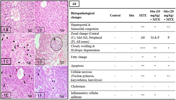
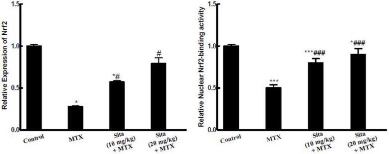
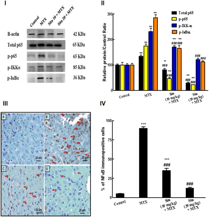
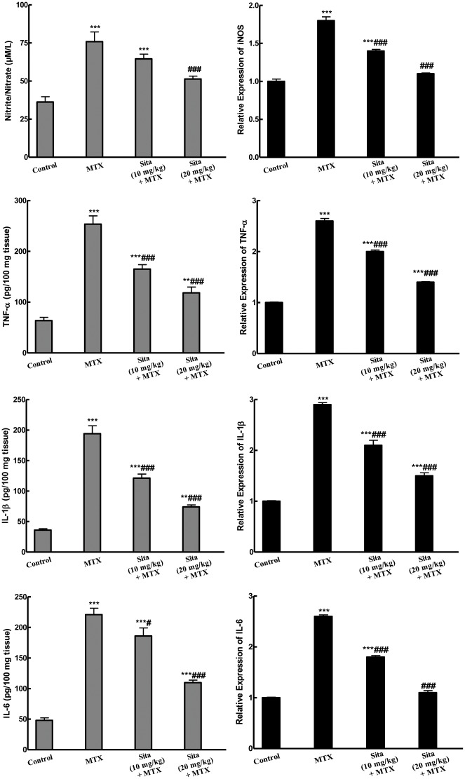
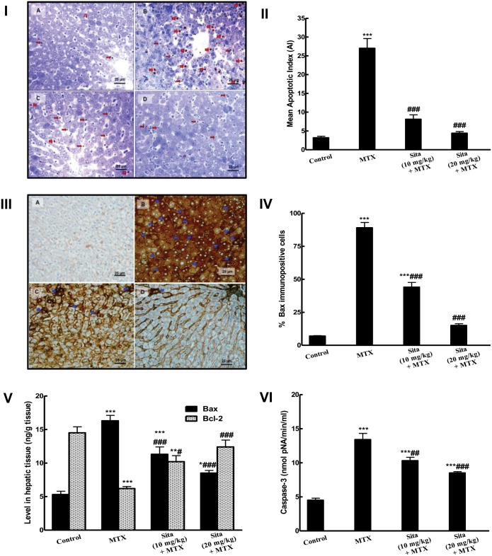
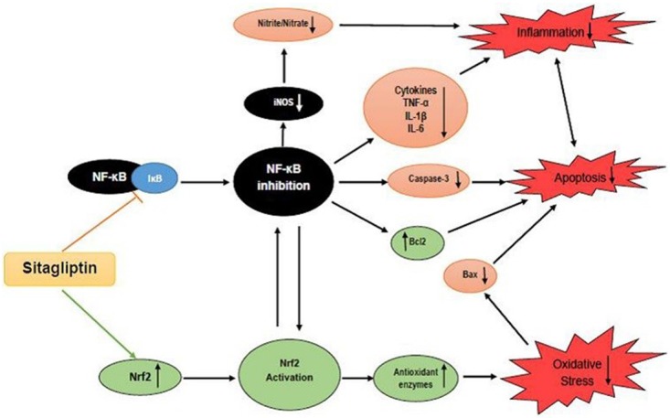
Sitagliptin is selective dipeptidyl peptidase-4 inhibitor (DPP4-I), used clinically as a new oral anti-diabetic agent. This study explored the underlying mechanisms of the hepatoprotective role of sitagliptin pretreatment against methotrexate (MTX) induced hepatotoxicity in mice. Forty mice were divided into four groups (10 mice each); control, MTX, and two sitagliptin groups (pretreated with sitagliptin 10 and 20 mg/kg/day, respectively) for five consecutive days prior to MTX injection. Results showed that MTX induced marked hepatic injury in the form of cloudy swelling, hydropic degeneration, apoptosis and focal necrosis in all hepatic zones. Biochemical analysis revealed significant increase in the serum transaminases, alkaline phosphatase and lactate dehydrogenase in MTX group. Oxidative stress and depressed antioxidant system of the hepatic tissues were evident in MTX group. MTX down-regulated nuclear factor erythroid 2-related factor 2 (Nrf2) expression and reduced its binding capacity. Additionally, MTX increased the activation and the expression of nuclear factor kappa-B (NF-κB) and downstream inflammatory mediators. MTX induced the activation of inducible nitric oxide synthase (iNOS) and increased nitrite/nitrate level. Finally, hepatic cellular apoptosis was clearly obvious in MTX-intoxicated animals using TUNEL staining. Also, there was increase in the immunoexpression of pro-apoptotic protein Bax, increase in Bax and caspase-3 levels and decrease in the level of anti-apoptotic Bcl2 in liver. On the other hand, sitagliptin pretreatment significantly ameliorated all of the above mentioned biochemical, histopathological, immunohistochemical changes induced by MTX. These results provide new evidences that the hepatoprotective effect of sitagliptin is possibly mediated through modulation of Nrf2 and NF-κB signaling pathways with subsequent suppression of inflammatory and apoptotic processes.


Conflict of interest statement

Competing Interests: The authors have declared that no competing interests exist.

Figures

Fig 1
Fig 1. Sitagliptin (Sita) pretreatment ameliorates Methotrexate (MTX)-induced histopathological lesions in liver of mice (H&E stain, 400×).
A. Control group showing normal liver histology, B. Sita group showing normal liver histology, C. MTX group showing cloudy swelling, hydropic degeneration (Chevron), fatty change (Arrow heads), shrinked apoptotic cells with pyknotic small nuclei and esinophic cytoplasm (wavy arrows) and focal necrosis (Arrows) in all hepatic zones. Many hepatocytes show signs of necrosis (Arrows) (pyknosis, kariorrhexis and karyolysis), D. Portal area of MTX group showed the periportal inflammatory cellular infiltrate (circle with star), E. Sita (10 mg/kg) + MTX group showing reduction in the histopathological lesions in the central hepatic lobular zone, F. Sita (20 mg/kg) + MTX group showing a significant reduction in the histopathological lesions in all hepatic lobular zones. G. Semi-quantitative scoring showing the hepatoprotective effects of Sita.
Fig 2
Fig 2. Sitagliptin (Sita) pretreatment improves Methotrexate (MTX)-induced reduction in Nrf2 expression and binding activity in the hepatic tissue of mice.
n = 10. *P < 0.05, ***P < 0.001 vs. the control. ###P < 0.001 vs. the MTX group (ANOVA followed by Tukey-Kramer multiple comparison).
Fig 3
Fig 3. Sitagliptin (Sita) pretreatment suppresses Methotrexate (MTX)-induced activation of nuclear factor kappa-B (NF-κB) pathway in the hepatic tissue of mice.
I. Western Blot for NF-κB p65, p-p65, p-IKKα, p-IκBα. II. Quantification of the protein intensities of relative protein level of NF-κB p65, p-p65, p-IKKα, p-IκBα. β-actin served as internal control. III. Immunohistochemical staining of nuclear NF-κB p65 (400×): A. Control group with focal light brown immunostaining, B. MTX group, showing intense brown immunostaining of NF-κB (Red arrows) (C, D) Sitagliptin + MTX groups, showing a marked decrease in the expression of NF-κB. IV. Semiquantitative analysis of NF-κB immunohistochemical staining results in liver tissues of different groups, expressed as % of NF-κB immunopositive cells across 10 different fields for each TmA section. n = 10. **P < 0.01, ***P < 0.001 vs. the control. ###P < 0.001 vs. the MTX group (ANOVA followed by Tukey-Kramer multiple comparison).
Fig 4
Fig 4. Sitagliptin (Sita) pretreatment alleviates Methotrexate (MTX)-induced increase in the levels and the expression of different inflammatory cytokines in the hepatic tissues of mice.
n = 10. **P < 0.01, ***P < 0.001 vs. the control. #P < 0.05, ###P < 0.001 vs. the MTX group (ANOVA followed by Tukey-Kramer multiple comparison).
Fig 5
Fig 5. Sitagliptin (Sita) pretreatment counteracts apoptosis and improves apoptotic markers in the hepatic tissues of Methotrexate (MTX)-treated mice.
I. TUNEL-positive apoptotic liver cells nuclei (Thick red arrow) and bodies (Thin red arrow) detected in the liver sections were: (A) Occasionally seen in Control mice group; (B) Frequently observed in mice group intoxicated with MTX; (C&D) Clearly decreased in sita pretreated groups with more marked reduction in the number of apoptotic cell and bodies seen in group pretreated with sita 20 mg/kg. II. Effect on mean apoptotic index (the number of TUNEL-positive cells/high- power field (× 400) across 10 different fields for each TmA section). III. The expression of pro-apoptotic marker Bax by immunohistochemical staining (400×): A) Control group with focal light brown cytoplasmic immunostaining in less than 1% of cells; B) MTX group, showing intense brown cytoplasmic immunostaining of Bax (Blue arrows) in more than 95% of cells (C, D) Sita + MTX groups, showing a marked decrease in the cytoplasmic expression of Bax in a dose dependent manner of sita. IV. Semiquantitative analysis of Bax immunohistochemical staining results in liver tissues of different groups, expressed as % of immunopositive cells in TmA sections of all animals of each group, 10 different fields/section. V. Levels of Bax and Bcl2 in hepatic tissue. VI. Caspase activity in hepatic tissue. n = 10. *P < 0.05, **P < 0.01, ***P < 0.001 vs. the control. #P < 0.05, ##P < 0.01, ###P < 0.001 vs. the MTX group (ANOVA followed by Tukey-Kramer multiple comparison).
Fig 6
Fig 6. Schematic diagram summarizing the possible mechanisms for the hepatoprotective effects of Sitagliptin (Sita) against Methotrexate (MTX)-induced hepatotoxicity.

References

    1. Hytiroglou P, Tobias H, Saxena R, Abramidou M, Papadimitriou CS, Theise ND (2004) The canals of hering might represent a target of methotrexate hepatic toxicity. Am J Clin Pathol 121: 324–329. 10.1309/5HR9-0TNC-4Q4J-RXWX - DOI - PubMed
    1. Dalaklioglu S, Genc GE, Aksoy NH, Akcit F, Gumuslu S (2013) Resveratrol ameliorates methotrexate-induced hepatotoxicity in rats via inhibition of lipid peroxidation. Hum Exp Toxicol 32: 662–671. 10.1177/0960327112468178 - DOI - PubMed
    1. Jahovic N, Cevik H, Sehirli AO, Yegen BC, Sener G (2003) Melatonin prevents methotraxate- induced hepatorenal oxidative injury in rats. J Pineal Res 34: 282–287. - PubMed
    1. Cetinkaya A, Bulbuloglu E, Kurutas EB, Kantarceken B (2006) N-acetylcycteine ameliorates methotrexate-induced oxidative liver damage in rats. Med Sci Monit 12: 274–278. - PubMed
    1. Uraz S, Tahan V, Aygun C, Eren F, Unluguzel G, Yuksel M, et al. (2008) Role of ursodeoxycholic acid in prevention of methotrexate-induced liver toxicity. Dig Dis Sci 53: 1071–1077. 10.1007/s10620-007-9949-3 - DOI - PubMed