

Inter-Regional Variations in Gene Expression and Age-Related Cortical Thinning in the Adolescent Brain
- PMID: 28334178
- PMCID: PMC6093352
- DOI: 10.1093/cercor/bhx040
Inter-Regional Variations in Gene Expression and Age-Related Cortical Thinning in the Adolescent Brain
Abstract
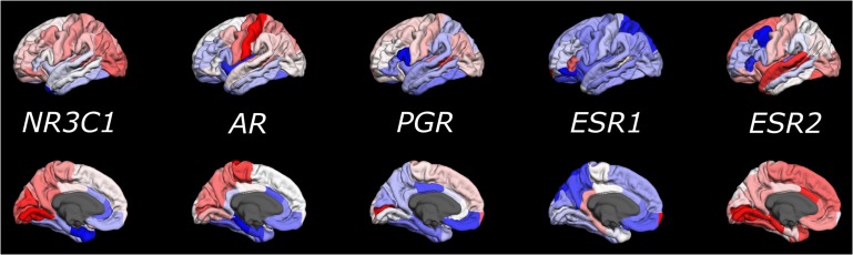
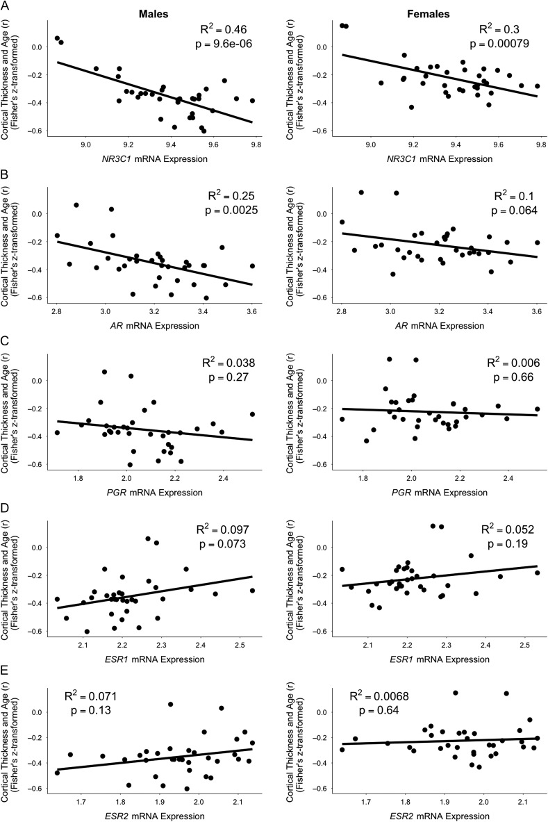
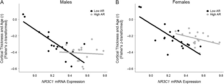
Age-related decreases in cortical thickness observed during adolescence may be related to fluctuations in sex and stress hormones. We examine this possibility by relating inter-regional variations in age-related cortical thinning (data from the Saguenay Youth Study) to inter-regional variations in expression levels of relevant genes (data from the Allen Human Brain Atlas); we focus on genes coding for glucocorticoid receptor (NR3C1), androgen receptor (AR), progesterone receptor (PGR), and estrogen receptors (ESR1 and ESR2). Across 34 cortical regions (Desikan-Killiany parcellation), age-related cortical thinning varied as a function of mRNA expression levels of NR3C1 in males (R2 = 0.46) and females (R2 = 0.30) and AR in males only (R2 = 0.25). Cortical thinning did not vary as a function of expression levels of PGR, ESR1, or ESR2 in either sex; this might be due to the observed low consistency of expression profiles of these 3 genes across donors. Inter-regional levels of the NR3C1 and AR expression interacted with each other vis-à-vis cortical thinning: age-related cortical thinning varied as a function of NR3C1 mRNA expression in brain regions with low (males: R2 = 0.64; females: R2 = 0.58) but not high (males: R2 = 0.0045; females: R2 = 0.15) levels of AR mRNA expression. These results suggest that glucocorticoid and androgen receptors contribute to cortical maturation during adolescence.
Figures
Similar articles
-
Corticosteroids and Regional Variations in Thickness of the Human Cerebral Cortex across the Lifespan.Cereb Cortex. 2020 Mar 21;30(2):575-586. doi: 10.1093/cercor/bhz108. Cereb Cortex. 2020. PMID: 31240317 Free PMC article.
-
Income inequality, gene expression, and brain maturation during adolescence.Sci Rep. 2017 Aug 7;7(1):7397. doi: 10.1038/s41598-017-07735-2. Sci Rep. 2017. PMID: 28784996 Free PMC article.
-
Sexually dimorphic androgen and estrogen receptor mRNA expression in the brain of túngara frogs.Horm Behav. 2010 Sep;58(4):619-27. doi: 10.1016/j.yhbeh.2010.06.013. Epub 2010 Jun 23. Horm Behav. 2010. PMID: 20600046
-
Accelerated longitudinal cortical thinning in adolescence.Neuroimage. 2015 Jan 1;104:138-45. doi: 10.1016/j.neuroimage.2014.10.005. Epub 2014 Oct 13. Neuroimage. 2015. PMID: 25312772
-
Polymorphisms in sex steroid receptors: From gene sequence to behavior.Front Neuroendocrinol. 2017 Oct;47:47-65. doi: 10.1016/j.yfrne.2017.07.003. Epub 2017 Jul 10. Front Neuroendocrinol. 2017. PMID: 28705582 Free PMC article. Review.
Cited by
-
Regional patterns of human cortex development correlate with underlying neurobiology.Nat Commun. 2024 Sep 12;15(1):7987. doi: 10.1038/s41467-024-52366-7. Nat Commun. 2024. PMID: 39284858 Free PMC article.
-
Maturation of the Human Cerebral Cortex During Adolescence: Myelin or Dendritic Arbor?Cereb Cortex. 2019 Jul 22;29(8):3351-3362. doi: 10.1093/cercor/bhy204. Cereb Cortex. 2019. PMID: 30169567 Free PMC article.
-
General and specific patterns of cortical gene expression as spatial correlates of complex cognitive functioning.Hum Brain Mapp. 2024 Mar;45(4):e26641. doi: 10.1002/hbm.26641. Hum Brain Mapp. 2024. PMID: 38488470 Free PMC article.
-
COVID-19 lockdown effects on adolescent brain structure suggest accelerated maturation that is more pronounced in females than in males.Proc Natl Acad Sci U S A. 2024 Sep 17;121(38):e2403200121. doi: 10.1073/pnas.2403200121. Epub 2024 Sep 9. Proc Natl Acad Sci U S A. 2024. PMID: 39250666 Free PMC article.
-
Life Event Stress and Reduced Cortical Thickness in Youth at Clinical High Risk for Psychosis and Healthy Control Subjects.Biol Psychiatry Cogn Neurosci Neuroimaging. 2022 Feb;7(2):171-179. doi: 10.1016/j.bpsc.2021.04.011. Epub 2021 Apr 28. Biol Psychiatry Cogn Neurosci Neuroimaging. 2022. PMID: 33930604 Free PMC article.
References
-
- Amlien IK, Fjell AM, Tamnes CK, Grydeland H, Krogsrud SK, Chaplin TA, Rosa MGP, Walhovd KB. 2016. Organizing principles of human cortical development – thickness and area from 4 to 30 years: insights from comparative primate neuroanatomy. Cereb Cortex. 26:257–267. - PubMed
-
- Cardinale F, Chinnici G, Bramerio M, Mai R, Sartori I, Cossu M, Lo Russo G, Castana L, Colombo N, Caborni C, et al. . 2014. Validation of freesurfer-estimated brain cortical thickness: comparison with histologic measurements. Neuroinformatics. 12:535–542. - PubMed
Publication types
MeSH terms
Substances
Grants and funding
LinkOut - more resources
Full Text Sources
Other Literature Sources
Medical
Research Materials
Miscellaneous
