. 2017 Apr 1;38(4):474-483.
doi: 10.1093/carcin/bgx023.

PPARα regulates tumor cell proliferation and senescence via a novel target gene carnitine palmitoyltransferase 1C

Affiliations

PPARα regulates tumor cell proliferation and senescence via a novel target gene carnitine palmitoyltransferase 1C

Yixin Chen et al. Carcinogenesis. .

Abstract

Carnitine palmitoyltransferase 1C (CPT1C), an enzyme located in the outer mitochondria membrane, has a crucial role in fatty acid transport and oxidation. It is also involved in cell proliferation and is a potential driver for cancer cell senescence. However, its upstream regulatory mechanism is unknown. Peroxisome proliferator activated receptor α (PPARα) is a ligand-activated transcription factor that regulates lipid metabolism and tumor progression. The current study aimed to elucidate whether and how PPARα regulates CPT1C and then affects cancer cell proliferation and senescence. Here, for the first time we report that PPARα directly activated CPT1C transcription and CPT1C was a novel target gene of PPARα, as revealed by dual-luciferase reporter and chromatin immunoprecipitation (ChIP) assays. Moreover, regulation of CPT1C by PPARα was p53-independent. We further confirmed that depletion of PPARα resulted in low CPT1C expression and then inhibited proliferation and induced senescence of MDA-MB-231 and PANC-1 tumor cell lines in a CPT1C-dependent manner, while forced PPARα overexpression promoted cell proliferation and reversed cellular senescence. Taken together, these results indicate that CPT1C is a novel PPARα target gene that regulates cancer cell proliferation and senescence. The PPARα-CPT1C axis may be a new target for the intervention of cancer cellular proliferation and senescence.


Figures

Figure 1.
Figure 1.
PPARα regulates CPT1C mRNA and protein expression. (A) RT-PCR analysis was used to determine the expression of PPARα and CPT1C in PANC-1 and MDA-MB-231 cells after down-regulating PPARα by siRNA or its inhibitor GW6471 (10 μM) and up-regulating PPARα with pGST-PPARα plasmid or its agonist WY14643 (100 μM). Data are the mean ± SEM (n = 4). (B) Western blot was used to measure protein level of PPARα and CPT1C as the same conditions described as above where GAPDH was used as a loading control. Gray scanning was evaluated by Quantity one. Data are the mean ± SEM (n = 3). (C) MDA-MB-231 cell line were stained for CPT1C by immunohistochemistry.
Figure 2.
Figure 2.
PPARα directly activates CPT1C transcription. (A) A series of dual-luciferase reporter gene assays were conducted in HEK-293T cells to compare reporter activities among plasmids with different lengths of CPT1C promoter regions. Data are the mean ± SEM (n = 6). (B) Dual-luciferase reporter gene assays results after integrating together. Data are the mean ± SEM (n = 6). (C) Four different PPRE regions called DRn were predicted in 3.0 kb CPT1C promoter region by bioinformatics. (D) MDA-MB-231 cells were treated with pGST-PPARα plasmid for 24 h then ChIP analysis was performed. DNA samples after precipitation reaction were purified and amplified through qPCR.
Figure 3.
Figure 3.
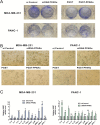
PPARα modulates cancer cell viability and proliferation. (A) WST-8 assay of MDA-MB-231 and PANC-1 cells were performed to examine cell viability after knocking down or overexpressing PPARα. (B) BrdU activity measured proliferation capacity of cells with treatment in siRNA PPARα or pGST-PPARα plasmid. (C) MDA-MB-231 cell lines were stained for Ki67 by means of immunohistochemistry. (D) Cell cycles of two cell lines were determined by flow cytometry when depletion of PPARα. Data are the mean ± SEM, P < 0.05 versus siControl or vehicle.
Figure 4.
Figure 4.
PPARα silencing induces cancer cell senescence. (A) Cells were stained with crystal violet for colony formation after being cultured for an additional 14 days. (B) SA-β-gal activity was measured to represent degree of senescence of tumor cells. (C) RT-PCR analysis was used to determine the expression of SASP factors such as IL-6, IL-7, IL-8, IL-1α, IL-1β, TNFα, TGFB1, ATM, MMP-1, MMP-3 and MCP-1 in two cell lines. Data are the mean ± SEM (n = 4).
Figure 5.
Figure 5.
PPARα regulates CPT1C expression in a p53 independent way. (A) RT-PCR analysis was used to determine the expression of PPARα and p53 mRNA levels in MDA-MB-231 cell line after down-regulating PPARα by siRNA or its inhibitor GW6471 (10 μM) and upregulating PPARα by overexpression plasmid pGST-PPARαor its agonist WY14643 (100 μM). Data are the mean ± SEM (n = 4). (B) Western blot was used to measure protein levels of PPARα and p53 as the same conditions as above by regarding GAPDH as internal references. Gray scanning was evaluated by Quantity one. Data are the mean ± SEM (n = 3). (C) RT-PCR analysis showed that the effect of PPARα on CPT1C expression still remained after silencing p53 expression. Data are the mean ± SEM (n = 4).

Similar articles

Cited by

References

    1. Sierra A.Y., et al. (2008) CPT1c is localized in endoplasmic reticulum of neurons and has carnitine palmitoyltransferase activity. J. Biol. Chem., 283, 6878–6885. - PubMed
    1. Bonnefont J.P., et al. (2004) Carnitine palmitoyltransferases 1 and 2: biochemical, molecular and medical aspects. Mol. Aspects Med., 25, 495–520. - PubMed
    1. Wolfgang M.J., et al. (2006) The brain-specific carnitine palmitoyltransferase-1c regulates energy homeostasis. Proc. Natl. Acad. Sci. USA, 103, 7282–7287. - PMC - PubMed
    1. Wolfgang M.J., et al. (2008) Brain-specific carnitine palmitoyl-transferase-1c: role in CNS fatty acid metabolism, food intake, and body weight. J. Neurochem., 105, 1550–1559. - PMC - PubMed
    1. Gao X.F., et al. (2009) Enhanced susceptibility of Cpt1c knockout mice to glucose intolerance induced by a high-fat diet involves elevated hepatic gluconeogenesis and decreased skeletal muscle glucose uptake. Diabetologia, 52, 912–920. - PubMed

Publication types