

LRIT3 Differentially Affects Connectivity and Synaptic Transmission of Cones to ON- and OFF-Bipolar Cells
- PMID: 28334377
- PMCID: PMC5374884
- DOI: 10.1167/iovs.16-20745
LRIT3 Differentially Affects Connectivity and Synaptic Transmission of Cones to ON- and OFF-Bipolar Cells
Abstract
Purpose: Mutations in LRIT3 lead to complete congenital stationary night blindness (cCSNB). Using a cCSNB mouse model lacking Lrit3 (nob6), we recently have shown that LRIT3 has a role in the correct localization of TRPM1 (transient receptor potential melastatin 1) to the dendritic tips of ON-bipolar cells (BCs), contacting both rod and cone photoreceptors. Furthermore, postsynaptic clustering of other mGluR6 cascade components is selectively eliminated at the dendritic tips of cone ON-BCs. The purpose of this study was to further define the role of LRIT3 in structural and functional organization of cone synapses.
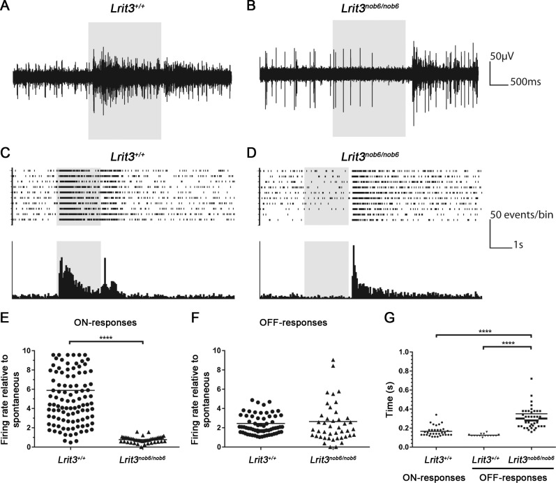
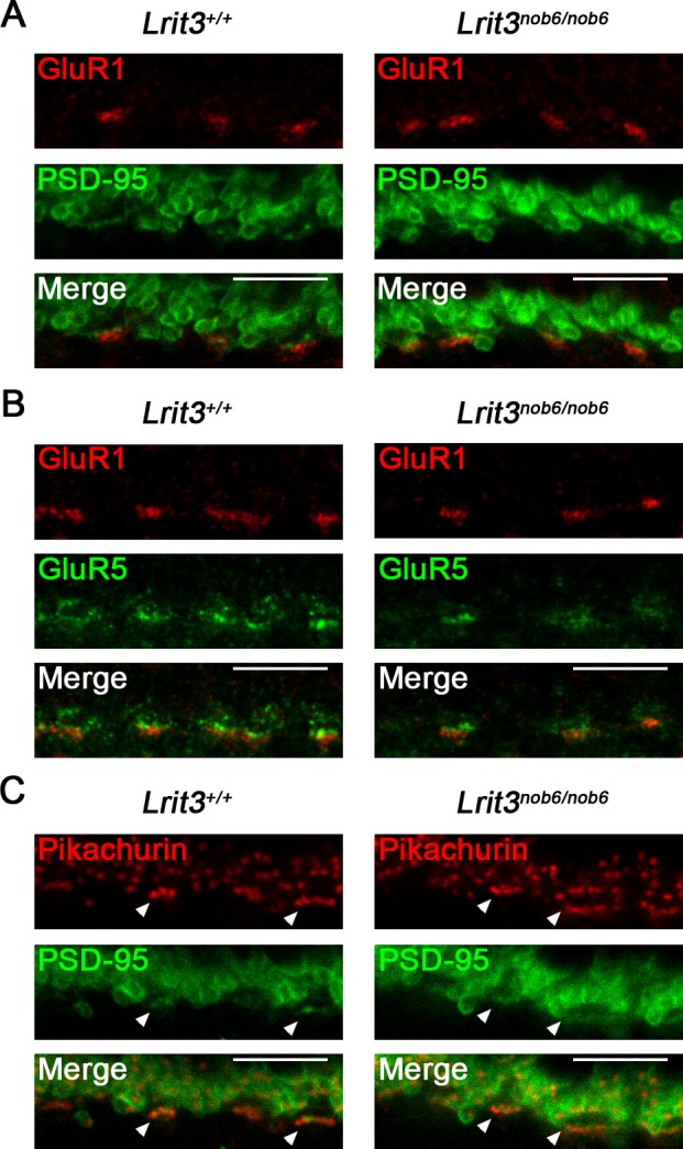
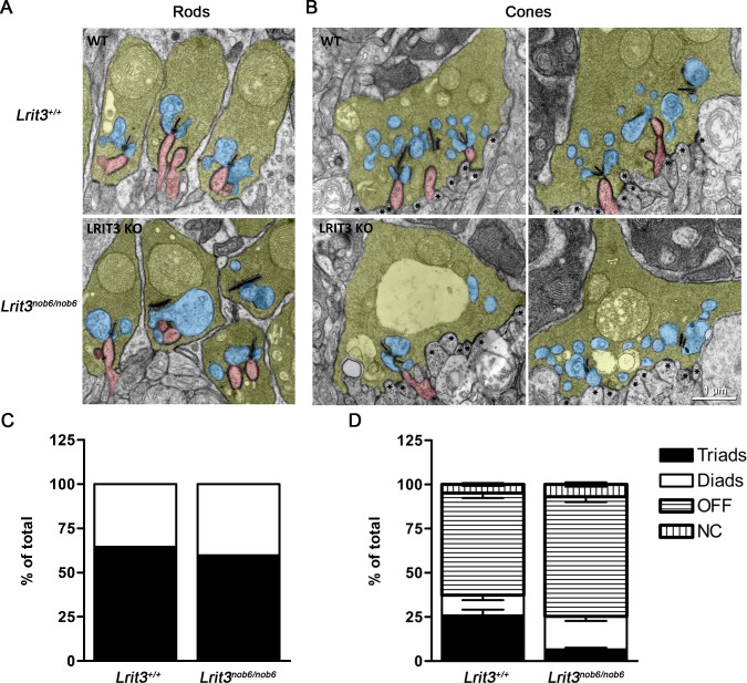
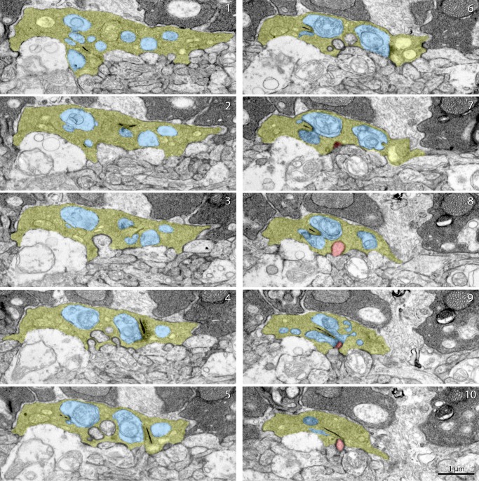
Methods: Exhaustive electroretinogram analysis was performed in a patient with LRIT3 mutations. Multielectrode array recordings were performed at the level of retinal ganglion cells in nob6 mice. Targeting of GluR1 and GluR5 at the dendritic tips of OFF-BCs in nob6 retinas was assessed by immunostaining and confocal microscopy. The ultrastructure of photoreceptor synapses was evaluated by electron microscopy in nob6 mice.
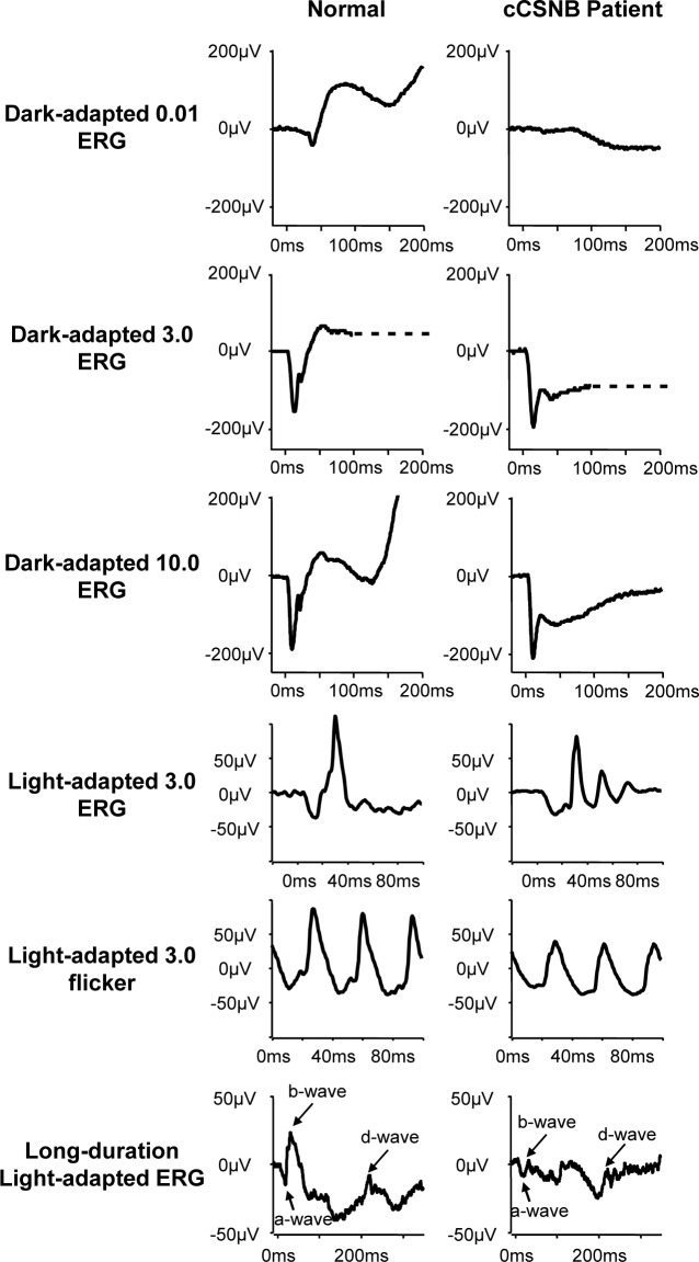
Results: The patient with LRIT3 mutations had a selective ON-BC dysfunction with relatively preserved OFF-BC responses. In nob6 mice, complete lack of ON-pathway function with robust, yet altered signaling processing in OFF-pathways was detected. Consistent with these observations, molecules essential for the OFF-BC signaling were normally targeted to the synapse. Finally, synaptic contacts made by ON-BC but not OFF-BC neurons with the cone pedicles were disorganized without ultrastructural alterations in cone terminals, horizontal cell processes, or synaptic ribbons.
Conclusions: These results suggest that LRIT3 is likely involved in coordination of the transsynaptic communication between cones and ON-BCs during synapse formation and function.
Figures
Similar articles
-
LRIT3 is essential to localize TRPM1 to the dendritic tips of depolarizing bipolar cells and may play a role in cone synapse formation.Eur J Neurosci. 2015 Aug;42(3):1966-75. doi: 10.1111/ejn.12959. Epub 2015 Jul 4. Eur J Neurosci. 2015. PMID: 25997951 Free PMC article.
-
Amyloid Precursor-Like Protein 2 deletion-induced retinal synaptopathy related to congenital stationary night blindness: structural, functional and molecular characteristics.Mol Brain. 2016 Jun 8;9(1):64. doi: 10.1186/s13041-016-0245-z. Mol Brain. 2016. PMID: 27267879 Free PMC article.
-
A Naturally Occurring Canine Model of Autosomal Recessive Congenital Stationary Night Blindness.PLoS One. 2015 Sep 14;10(9):e0137072. doi: 10.1371/journal.pone.0137072. eCollection 2015. PLoS One. 2015. PMID: 26368928 Free PMC article.
-
Properties and functions of TRPM1 channels in the dendritic tips of retinal ON-bipolar cells.Eur J Cell Biol. 2015 Jul-Sep;94(7-9):420-7. doi: 10.1016/j.ejcb.2015.06.005. Epub 2015 Jun 3. Eur J Cell Biol. 2015. PMID: 26111660 Review.
-
Shedding light on myopia by studying complete congenital stationary night blindness.Prog Retin Eye Res. 2023 Mar;93:101155. doi: 10.1016/j.preteyeres.2022.101155. Epub 2023 Jan 19. Prog Retin Eye Res. 2023. PMID: 36669906 Review.
Cited by
-
Expression and distribution of trophoblast glycoprotein in the mouse retina.J Comp Neurol. 2020 Jul;528(10):1660-1671. doi: 10.1002/cne.24850. Epub 2020 Jan 6. J Comp Neurol. 2020. PMID: 31891182 Free PMC article.
-
Characterizing Retinal Sensitivity and Structure in Congenital Stationary Night Blindness: A Combined Microperimetry and OCT Study.Invest Ophthalmol Vis Sci. 2024 Jun 3;65(6):35. doi: 10.1167/iovs.65.6.35. Invest Ophthalmol Vis Sci. 2024. PMID: 38916884 Free PMC article.
-
LRIT3 is Required for Nyctalopin Expression and Normal ON and OFF Pathway Signaling in the Retina.eNeuro. 2020 Feb 11;7(1):ENEURO.0002-20.2020. doi: 10.1523/ENEURO.0002-20.2020. Print 2020 Jan/Feb. eNeuro. 2020. PMID: 31959619 Free PMC article.
-
Efficient in vivo labeling of endogenous proteins with SMART delineates retina cellular and synaptic organization.Nat Commun. 2025 Apr 22;16(1):3768. doi: 10.1038/s41467-025-58945-6. Nat Commun. 2025. PMID: 40263339 Free PMC article.
-
Molecular mechanisms underlying selective synapse formation of vertebrate retinal photoreceptor cells.Cell Mol Life Sci. 2020 Apr;77(7):1251-1266. doi: 10.1007/s00018-019-03324-w. Epub 2019 Oct 4. Cell Mol Life Sci. 2020. PMID: 31586239 Free PMC article. Review.
References
-
- Nakajima Y,, Iwakabe H,, Akazawa C,, et al. Molecular characterization of a novel retinal metabotropic glutamate receptor mGluR6 with a high agonist selectivity for L-2-amino-4-phosphonobutyrate. J Biol Chem. 1993; 268: 11868–11873. - PubMed
-
- Nomura A,, Shigemoto R,, Nakamura Y,, Okamoto N,, Mizuno N,, Nakanishi S. Developmentally regulated postsynaptic localization of a metabotropic glutamate receptor in rat rod bipolar cells. Cell. 1994; 77: 361–369. - PubMed
-
- Masu M,, Iwakabe H,, Tagawa Y,, et al. Specific deficit of the ON response in visual transmission by targeted disruption of the mGluR6 gene. Cell. 1995; 80: 757–765. - PubMed
Publication types
MeSH terms
Substances
Supplementary concepts
Grants and funding
LinkOut - more resources
Full Text Sources
Other Literature Sources
Molecular Biology Databases
Miscellaneous
