. 2017 Mar 22;93(6):1325-1333.e3.
doi: 10.1016/j.neuron.2017.02.043.

Gradual Suppression of Transcytosis Governs Functional Blood-Retinal Barrier Formation

Affiliations

Gradual Suppression of Transcytosis Governs Functional Blood-Retinal Barrier Formation

Brian Wai Chow et al. Neuron. .

Abstract

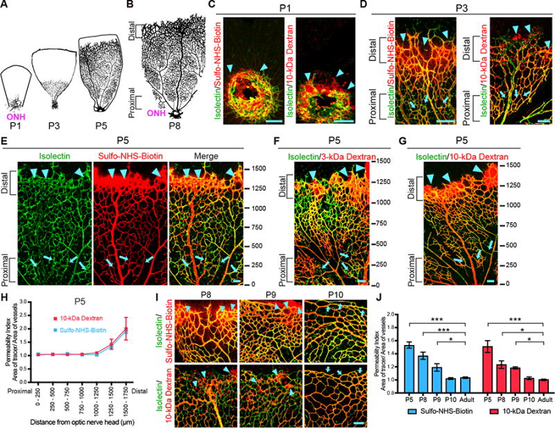
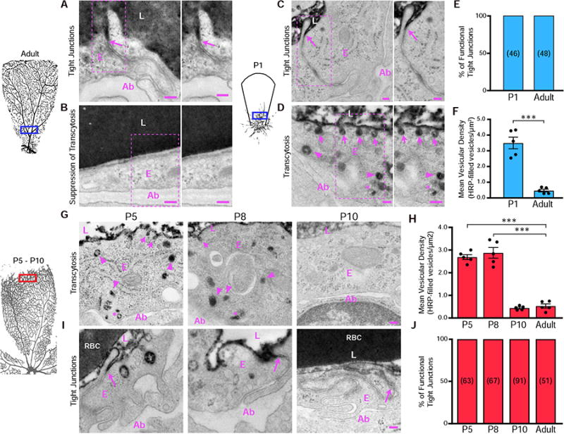
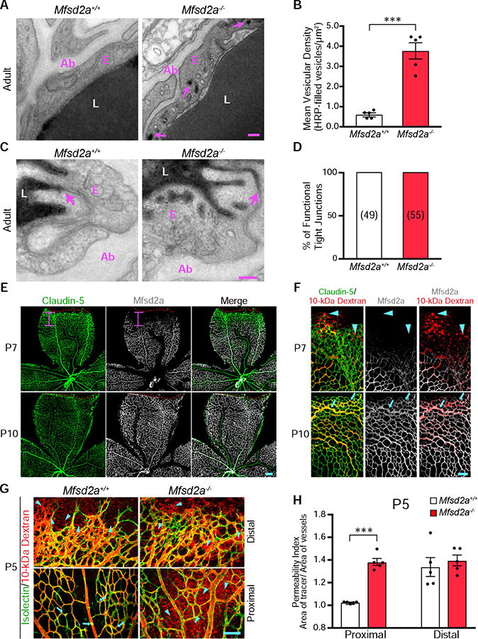
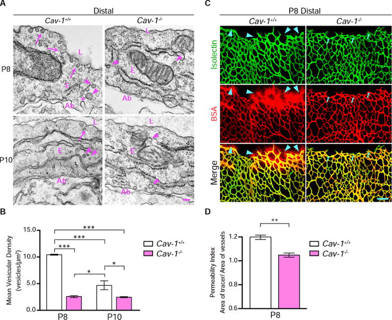
Blood-central nervous system (CNS) barriers partition neural tissues from the blood, providing a homeostatic environment for proper neural function. The endothelial cells that form blood-CNS barriers have specialized tight junctions and low rates of transcytosis to limit the flux of substances between blood and CNS. However, the relative contributions of these properties to CNS barrier permeability are unknown. Here, by studying functional blood-retinal barrier (BRB) formation in mice, we found that immature vessel leakage occurs entirely through transcytosis, as specialized tight junctions are functional as early as vessel entry into the CNS. A functional barrier forms only when transcytosis is gradually suppressed during development. Mutant mice with elevated or reduced levels of transcytosis have delayed or precocious sealing of the BRB, respectively. Therefore, the temporal regulation of transcytosis governs the development of a functional BRB, and suppression of transcytosis is a principal contributor for functional barrier formation.

Keywords: Cav-1; Mfsd2a; blood-CNS barrier; blood-brain barrier; blood-retinal barrier; endothelial cells; pericytes; retinal vasculature; tight junctions; transcytosis.


Figures

Figure 1
Figure 1. Spatio-temporal characterization of functional BRB formation, See also Figure S1, S2 and S5
Intravenous injections of tracers in neonatal mice reveal that functional BRB is formed gradually from proximal to distal in the primary plexus. (A-B) Spatio-temporal development of the mouse retina primary plexus. (A)Vessel ingression at the optic nerve head (ONH) occurs at P1. Vessels then expand from the ONH to the retinal periphery during postnatal development. (B) Vessels reach the peripheral edge of the retina at P8. A spatio-temporal gradient of vessel maturation is observed: the vessels proximal to the ONH are more mature whereas the vessels distal to the ONH are nascent. (C) In P1 retinas, when blood vessels (isolectin; green) first enter the retina, Sulfo-NHS-Biotin (left) and 10-kDa dextran (right) tracer (red) leakage is observed around budding vessels (arrowheads). (D-G) In P3 (D) and P5 (E-G) retinas, a gradient of barrier functionality is observed. More mature vessels proximal to the ONH confine tracer (arrows) whereas nascent vessels distal to the ONH leak tracer (arrowheads). Ticks in (E-G) represent distance in microns from the ONH. 0 is slightly before the ONH. (H) Permeability index of P5 retinas from tracer-injected pups reveals the functional BRB is gradually formed in a proximal to distal fashion. The permeability index is measured by the ratio of tracer-positive area over isolectin-positive area. A ratio greater than 1 indicates that the BRB is permeable and immature. A ratio of 1 indicates that the BRB is impermeable and hence mature. 250 μm2 field of views were sampled and tiled starting from 0 to the angiogenic front. The average distance from the ONH to the angiogenic front at P5 is 1651 ± 288 μm. Data are mean ± s.e.m. (n = 5-6). (I) Higher magnification of distal vessels of the primary plexus show Sulfo-NHS-Biotin (top) and 10-kDa Dextran (bottom) tracer leakage at P8 and P9 (arrowheads). At P10, both tracers are confined in distal vessels (arrows). (J) Quantification of distal vessel permeability for Sulfo-NHS-Biotin and 10-kDa dextran throughout BRB development. Data are mean ± s.e.m. (n = 5-6 mice per age per tracer group, from 3 different litters). Statistical significance was determined by oneway ANOVA with a post-hoc Bonferroni multiple comparison adjustment, comparing the various neonatal ages with the adult in the respective Sulfo-NHS-Biotin and 10-kDa Dextran group. *, P < 0.05, ***, P < 0.001. Scale bar represents 100 μm for all panels.
Figure 2
Figure 2. Gradual suppression of transcytosis governs the development of an impermeable, functional BRB. See also Figure S3
As early as P1, budding CNS endothelial cells possess functional tight junctions but display bulk transcytosis. (A and B) EM of endothelial cells in the proximal retina of an HRP-injected adult mouse. (A) Specialized tight junctions are functional and prevent electron dense 3-3' diaminobenzidine (DAB) reaction product in the lumen from invading through the intercellular cleft (arrows). (B) Transcytosis was suppressed in endothelial cells as seen by negligible numbers of tracer-filled vesicles. Also shown is a magnification of the boxed areas. (C and D) EM of endothelial cells in the proximal retina from an HRP-injected P1 pup. (C) Tracer invades through the intracellular cleft between the endothelial cells but stops at junctions without invading to the abluminal side (arrows). (D) DAB reaction product filled the vesicles attached to the luminal membrane (arrows), in the cytoplasm (arrowheads) and near the abluminal membrane (asterisk). Also shown is a magnification of the boxed areas. (E) The percentage of functional tight junctions from the EM images (n = 5 mice per age; 15-20 vessels analyzed per mouse; number of tight junctions analyzed are displayed in parenthesis). (F) Number of tracer-filled vesicles in endothelial cells in P1 and adult mice. Data are presented as mean ± s.e.m. (n = 5 mice per age, each circle represents the average vesicular density from 15 – 20 vessels per mouse). Statistical significance was assessed by unpaired t-test. L – lumen, E – endothelial cell, Ab – abluminal. **, P < 0.01. Scale bar represents 100 nm in all panels. (G) At P5 and P8, many tracer-filled vesicles are observed at the luminal membrane (arrows), cytoplasm (arrowheads) and at the abluminal membrane (*) in distal endothelial cells. At P10, distal endothelial cells contain negligible numbers of tracer-filled vesicles. (H) Number of tracer-filled vesicular densities in distal vessels at P5, P8 and P10. Data are shown as mean ± s.e.m. (n= 5 mice per age; each circle represents the average vesicular density from 15 – 20 vessels per mouse. Statistical significance was determined by one-way ANOVA with a post-hoc Bonferroni multiple comparison adjustment, comparing distal vessels of the various neonatal ages to distal vessels of adults. (I) EM of distal vessels from P5, P8 and P10 retinas reveals tracer product halts at tight junctions (arrows) at all ages. (J) Percentage of functional tight junctions from distal vessels. (n = 5 mice per age; 15-20 vessels analyzed per mouse; number of tight junctions analyzed are displayed in the bars). L – lumen, E – endothelial cells, Ab – abluminal, RBC- red blood cell. ***, P < 0.001. Scale bar represents 100 nm in all panels.
Figure 3
Figure 3. Elevated levels of transcytosis deter functional BRB formation. See also Figure S4
(A) Transcytosis is increased in Mfsd2a−/&minus mice. Many tracer-filled vesicles (arrows) were observed in adult retinal endothelial cells from Mfsd2a−/&minus but not Mfsd2a+/+ mice. (B) Quantification of HRP-filled vesicles in retinal endothelial cells from adult Mfsd2a+/+ and Mfsd2a−/&minus mice. Data are shown as mean ± s.e.m. (n= 5 mice per genotype; each circle represents the average vesicular density from 18 – 20 vessels per mouse). Statistical significance was determined by unpaired t-test. (C) EM of the adult retina confirms that specialized tight junctions halt tracer product in both Mfsd2a+/+ and Mfsd2a−/&minus adult mice (D) Quantification of functional tight junctions from Mfsd2a+/+ and Mfsd2a−/&minus adult mice (n = 5 mice per genotype; 18-20 vessels analyzed per mouse; number of tight junctions analyzed are displayed in parenthesis). (E) Immunostaining for Claudin-5 (green) and Mfsd2a (white) on P7 and P10 retinas shows the lack of Mfsd2a expression in nascent, distal vessels at P7. The red dash lines indicate the angiogenic front as determined by Claudin-5 expression, and the pink bar indicates the length from the angiogenic front to the appearance of Mfsd2a expression. In contrast to P7, Mfsd2a is expressed in the distal vessels at P10 (n= 5 mice per age). (F) Mfsd2a expression correlates with functional BRB formation. Immunostaining for Claudin-5 and Mfsd2a in retinas from 10-kDa dextran injected P7 and P10 mice shows that at P7, extravasation of tracer (arrowheads) occurs at nascent vessels where Mfsd2a is absent. At P10, tracer is confined (arrows) in distal vessels where Mfsd2a is expressed (n=5 mice per age). (G) Genetic ablation of Mfsd2a results in incomplete formation of the functional BRB. After intravenous injection of 10-kDa dextran in P5 Mfsd2a+/+ and Mfsd2a−/&minus pups, tracer was confined (arrow) in proximal vessels (bottom) but leaked from distal vessels (top) in Mfsd2a+/+ mice (arrowheads). In contrast, tracer leaked into the retinal parenchyma from both proximal and distal vessels in Mfsd2a−/&minus mice (arrowheads). (H) Permeability index from proximal and distal regions of P5 Mfsd2a−/&minus and Mfsd2a+/+ littermates. Data are shown as mean ± s.e.m. (n=5 mice per genotype; each circle represents the average permeability from each mouse). Statistical significance was determined by unpaired t-test. ***, P < 0.001. Scale bars represents 100 nm in (A and C) and 100 μm in (E,F, and G).
Figure 4
Figure 4. Precocious suppression of transcytosis accelerates functional BRB formation
(A) Precocious suppression of transcytosis is observed in Cav-1−/&minus retinas. EM in distal vessels at P8 (top) reveals many vesicles associated with luminal (arrows) and abluminal membranes (*) and in the cytoplasm (arrowheads) in Cav-1+/+ endothelial cells. Cav-1−/&minus endothelial cells have drastically reduced numbers of vesicles. At P10 (bottom), few vesicles are observed in both Cav-1+/+ and Cav-1−/&minus distal vessels. (B) Quantifications of vesicles in distal vessels from Cav-1+/+ and Cav-1−/&minus mice at P8 and P10. Data are presented as mean ± s.e.m. (n= 3 mice per age per genotype, 15-20 vessels analyzed per mouse). Statistical significance was determined by two-way ANOVA with a post-hoc Bonferroni multiple comparison adjustment. (C) Intravenous injection of BSA (red) in P8 pups reveals tracer leakage in the retinal parenchyma from distal vessels of Cav-1+/+ (arrowheads) but not Cav-1−/&minus mice. (D) Permeability index from distal regions of P8 Cav-1+/+ and Cav-1−/&minus littermates. Data are presented as mean ± s.e.m. (n=3-4 mice per genotype). Statistical significance was determined by unpaired t-test. *, P < 0.05; **, P < 0.01; ***, P < 0.001. L – lumen, E – endothelial cells, Ab – abluminal. Scale bar represents 100 nm in (A) and 100 μm in (C).

Comment in

References

    1. Andreone BJ, Lacoste B, Gu C. Neuronal and Vascular Interactions Annu Rev Neurosci. 2015;38:25–46. - PMC - PubMed
    1. Armulik A, Genové G, Mäe M, Nisancioglu MH, Wallgard E, Niaudet C, He L, Norlin J, Lindblom P, Strittmatter K, Johansson BR, Betsholtz C. Pericytes regulate the blood-brain barrier. Nature. 2010;468:557–561. - PubMed
    1. Banks WA. From blood-brain barrier to blood-brain interface: new opportunities for CNS drug delivery. Nat Rev Drug Discov 2016 - PubMed
    1. Bell RD, Winkler EA, Sagare AP, Singh I, LaRue B, Deane R, Zlokovic BV. Pericytes Control Key Neurovascular Functions and Neuronal Phenotype in the Adult Brain and during Brain Aging. Neuron. 2010;68:409–427. - PMC - PubMed
    1. Ben-Zvi A, Lacoste B, Kur E, Andreone BJ, Mayshar Y, Yan H, Gu C. Mfsd2a is critical for the formation and function of the blood-brain barrier. Nature. 2014;509:507–511. - PMC - PubMed