How the embryonic chick brain twists
- PMID: 28334695
- PMCID: PMC5134006
- DOI: 10.1098/rsif.2016.0395
How the embryonic chick brain twists
Abstract
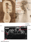
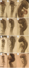
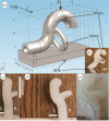
During early development, the tubular embryonic chick brain undergoes a combination of progressive ventral bending and rightward torsion, one of the earliest organ-level left-right asymmetry events in development. Existing evidence suggests that bending is caused by differential growth, but the mechanism for the predominantly rightward torsion of the embryonic brain tube remains poorly understood. Here, we show through a combination of in vitro experiments, a physical model of the embryonic morphology and mechanics analysis that the vitelline membrane (VM) exerts an external load on the brain that drives torsion. Our theoretical analysis showed that the force is of the order of 10 micronewtons. We also designed an experiment to use fluid surface tension to replace the mechanical role of the VM, and the estimated magnitude of the force owing to surface tension was shown to be consistent with the above theoretical analysis. We further discovered that the asymmetry of the looping heart determines the chirality of the twisted brain via physical mechanisms, demonstrating the mechanical transfer of left-right asymmetry between organs. Our experiments also implied that brain flexure is a necessary condition for torsion. Our work clarifies the mechanical origin of torsion and the development of left-right asymmetry in the early embryonic brain.
Keywords: axial rotation; biomechanics; embryonic development; left–right asymmetry; torsion.
© 2016 The Author(s).
Figures


Similar articles
-
Probing the Roles of Physical Forces in Early Chick Embryonic Morphogenesis.J Vis Exp. 2018 Jun 5;(136):57150. doi: 10.3791/57150. J Vis Exp. 2018. PMID: 29939170 Free PMC article.
-
On rotation, torsion, lateralization, and handedness of the embryonic heart loop: new insights from a simulation model for the heart loop of chick embryos.Anat Rec A Discov Mol Cell Evol Biol. 2004 May;278(1):481-92. doi: 10.1002/ar.a.20036. Anat Rec A Discov Mol Cell Evol Biol. 2004. PMID: 15103744
-
The role of mechanical forces in dextral rotation during cardiac looping in the chick embryo.Dev Biol. 2004 Aug 15;272(2):339-50. doi: 10.1016/j.ydbio.2004.04.033. Dev Biol. 2004. PMID: 15282152
-
Avian models and the study of invariant asymmetry: how the chicken and the egg taught us to tell right from left.Int J Dev Biol. 2018;62(1-2-3):63-77. doi: 10.1387/ijdb.180047ml. Int J Dev Biol. 2018. PMID: 29616741 Review.
-
Left-right asymmetry in heart development and disease: forming the right loop.Development. 2018 Nov 22;145(22):dev162776. doi: 10.1242/dev.162776. Development. 2018. PMID: 30467108 Review.
Cited by
-
Generation, Transmission, and Regulation of Mechanical Forces in Embryonic Morphogenesis.Small. 2022 Feb;18(6):e2103466. doi: 10.1002/smll.202103466. Epub 2021 Nov 26. Small. 2022. PMID: 34837328 Free PMC article. Review.
-
Alteration of mechanical stresses in the murine brain by age and hemorrhagic stroke.PNAS Nexus. 2024 Apr 24;3(4):pgae141. doi: 10.1093/pnasnexus/pgae141. eCollection 2024 Apr. PNAS Nexus. 2024. PMID: 38659974 Free PMC article.
-
Probing the Roles of Physical Forces in Early Chick Embryonic Morphogenesis.J Vis Exp. 2018 Jun 5;(136):57150. doi: 10.3791/57150. J Vis Exp. 2018. PMID: 29939170 Free PMC article.
-
Anatomical and Embryological Development of the Chick Cerebrum in Different Embryonic Periods.Vet Med Sci. 2025 Jan;11(1):e70124. doi: 10.1002/vms3.70124. Vet Med Sci. 2025. PMID: 39792061 Free PMC article.
References
-
- Taber LA. 1995. Biomechanics of growth, remodeling, and morphogenesis. Appl. Mech. Rev. 48, 487–545. (10.1115/1.3005109) - DOI
-
- Thompson DW. 1971. On growth and form. Cambridge, UK: Cambridge University Press.
Publication types
MeSH terms
Grants and funding
LinkOut - more resources
Full Text Sources
Other Literature Sources

