. 2017 Jun 2;45(10):6023-6036.
doi: 10.1093/nar/gkx135.

N1-methyl-pseudouridine in mRNA enhances translation through eIF2α-dependent and independent mechanisms by increasing ribosome density

Affiliations

N1-methyl-pseudouridine in mRNA enhances translation through eIF2α-dependent and independent mechanisms by increasing ribosome density

Yuri V Svitkin et al. Nucleic Acids Res. .

Abstract

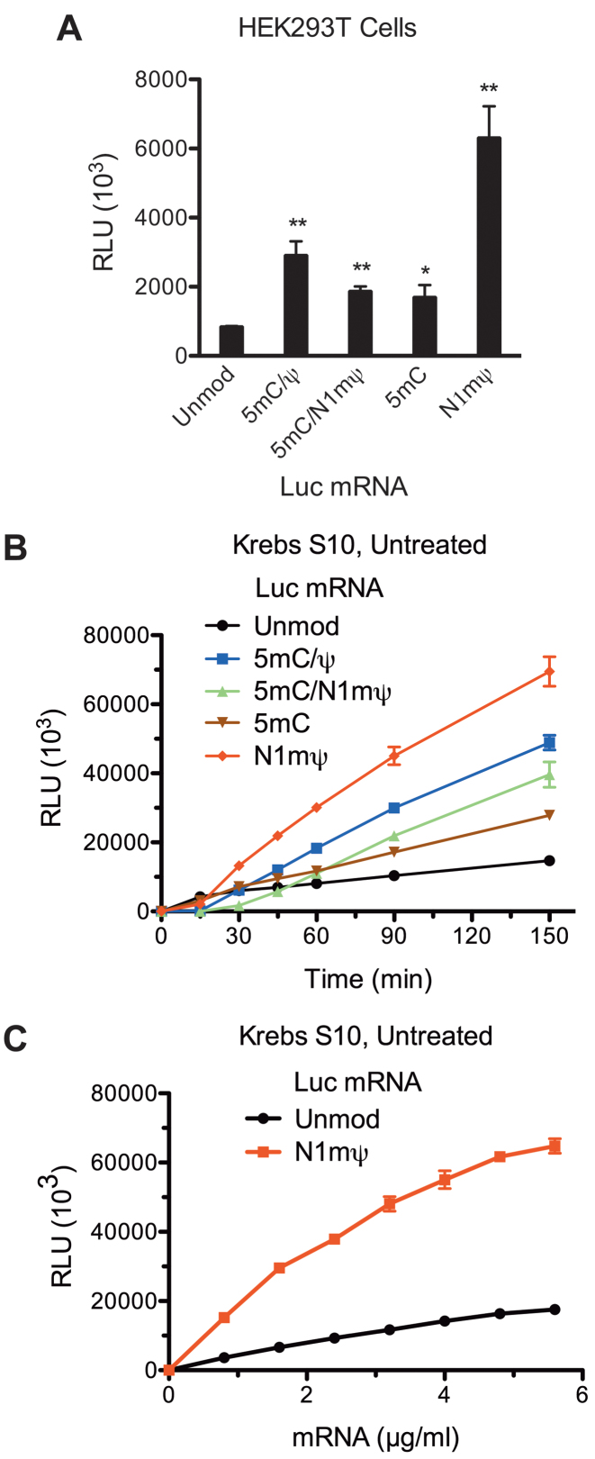
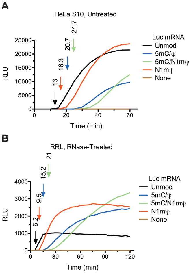
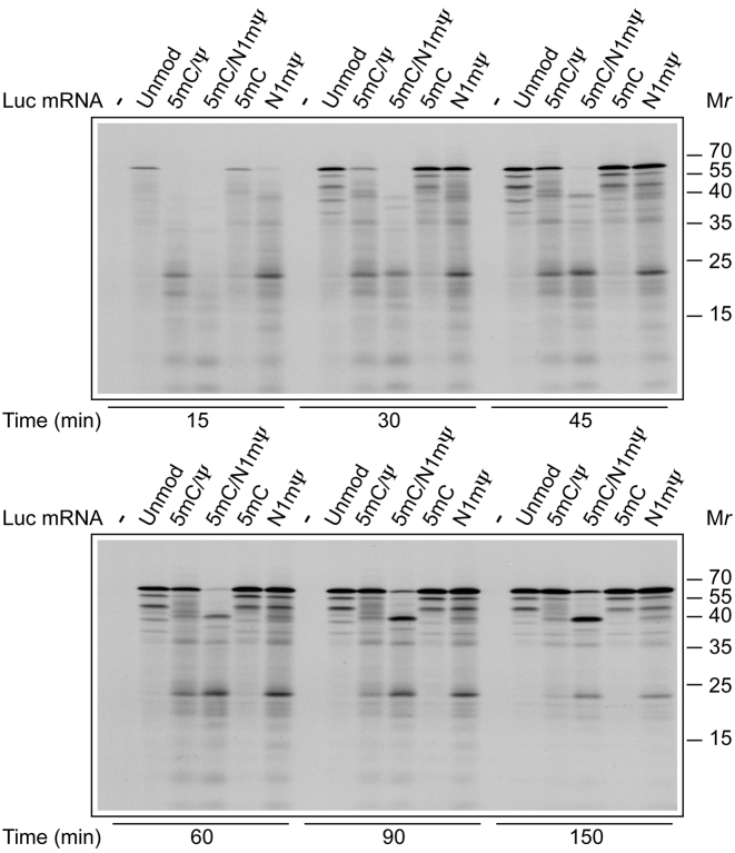
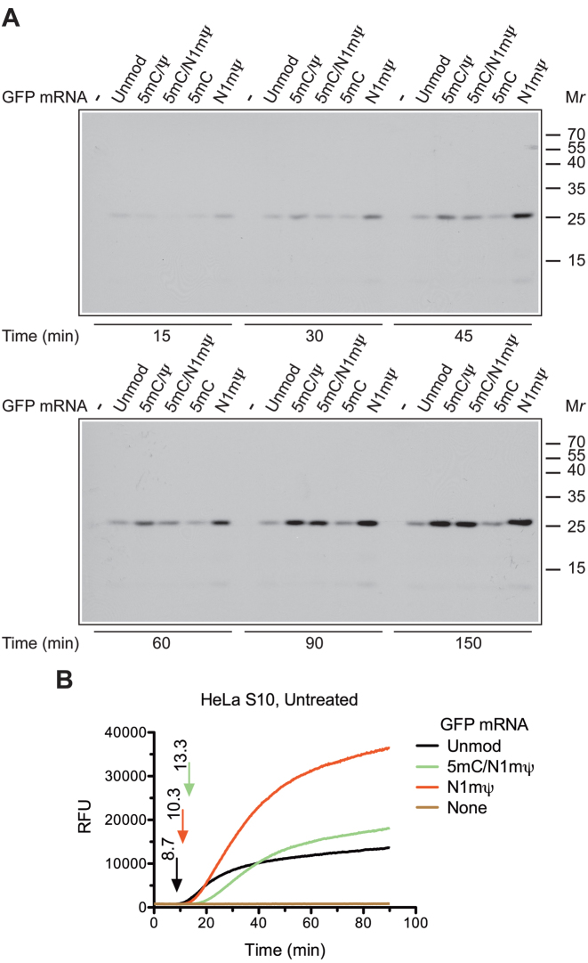
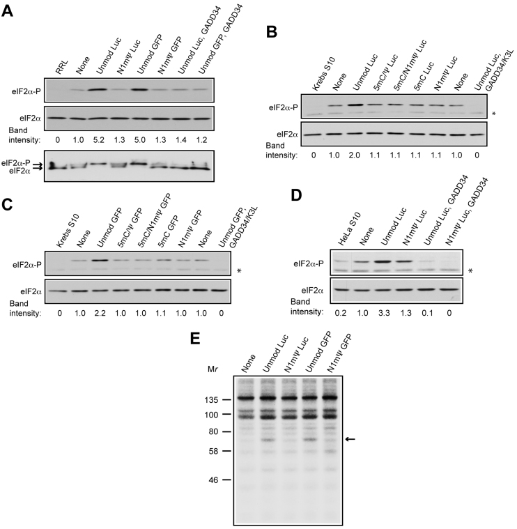
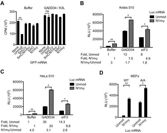
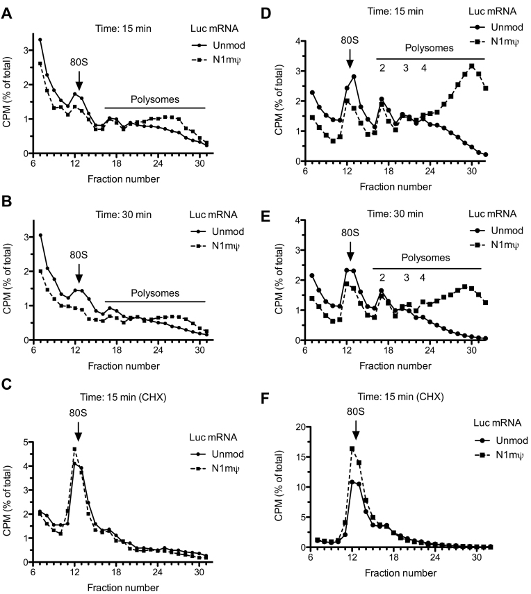
Certain chemical modifications confer increased stability and low immunogenicity to in vitro transcribed mRNAs, thereby facilitating expression of therapeutically important proteins. Here, we demonstrate that N1-methyl-pseudouridine (N1mΨ) outperforms several other nucleoside modifications and their combinations in terms of translation capacity. Through extensive analysis of various modified transcripts in cell-free translation systems, we deconvolute the different components of the effect on protein expression independent of mRNA stability mechanisms. We show that in addition to turning off the immune/eIF2α phosphorylation-dependent inhibition of translation, the incorporated N1mΨ nucleotides dramatically alter the dynamics of the translation process by increasing ribosome pausing and density on the mRNA. Our results indicate that the increased ribosome loading of modified mRNAs renders them more permissive for initiation by favoring either ribosome recycling on the same mRNA or de novo ribosome recruitment.


Figures

Figure 1.
Figure 1.
Recapitulation of the translational enhancement by modified nucleosides in mRNA in Krebs extract. (A) Nucleoside modifications conferring enhanced translation to Luc mRNA in cells. Luc mRNAs, either not containing (Unmod) or containing the 5 mC/Ψ, 5 mC/N1mΨ (N1-methyl-pseudouridine), 5 mC and N1mΨ nucleoside modifications, were transfected into HEK293T cells. Cells were lysed 4.5 h after transfection and luc activity was measured in 1% aliquots of the lysates. (B) Time course analysis of luc synthesis in untreated Krebs extracts supplemented with unmodified or 5 mC/Ψ, 5 mC/N1mΨ, 5 mC and N1mΨ-incorporated Luc mRNAs (4 μg/ml). At the indicated time points after beginning of translation at 30°C, 1-μl aliquots of the reaction mixtures were assayed for luc activity. (C) Unmodified and N1mΨ-modified Luc mRNA dose response of translation in Krebs extract. Luc and N1mΨ–Luc mRNAs were translated in untreated Krebs extracts at the indicated concentrations. Following incubation at 30°C for 4 h, 1-μl aliquots of the translation mixtures were assayed for luc activity. Data are means from three assays ± SD (*P < 0.05, **P < 0.01). Relative luciferase units (RLU).
Figure 2.
Figure 2.
Kinetics of luc synthesis in cell-free extracts translating unmodified or modified Luc mRNAs as determined by real-time measurements of Luc activity. Unmodified or 5 mC/Ψ, 5 mC/N1mΨ and N1mΨ-incorporated Luc mRNAs (20 μg/ml) were translated in untreated HeLa S10 extract (A) or RNase-treated RRL (B). The representative kinetic curves of luc synthesis and the background levels (None) are shown. The first time point at which the recorded signal is significantly above the background are taken as the durations of single translation cycle of mRNAs (indicated by arrows in matching colors). For details, see ‘Materials and Methods’ section.
Figure 3.
Figure 3.
Time course of synthesis of polypeptides in Krebs extracts programmed with unmodified or modified Luc mRNAs. Unmodified or 5 mC/Ψ, 5 mC/N1mΨ, 5 mC and N1mΨ-incorporated Luc mRNAs (4 μg/ml) were translated in RNase-treated Krebs extracts in the presence of 35S-methionine. At the indicated time points, aliquots of the reaction mixtures were withdrawn and fixed with SDS-sample buffer. Translation products were analyzed by SDS-PAGE and autoradiography. Molecular mass markers are indicated on the right.
Figure 4.
Figure 4.
Time course of GFP synthesis in cell-free extracts translating unmodified or modified GFP mRNAs. (A) Unmodified or 5 mC/Ψ, 5 mC/N1mΨ, 5 mC and N1mΨ-incorporated GFP mRNAs (4 μg/ml) were translated in RNase-treated Krebs extracts in the presence of 35S-methionine. At the indicated time points, aliquots of the reaction mixtures were withdrawn for analysis by SDS-PAGE and autoradiography. The positions of molecular mass markers are indicated on the right. (B) Real-time kinetic analysis of active GFP synthesis in untreated HeLa S10 extract programmed with unmodified or 5 mC/N1mΨ and N1mΨ-incorporated GFP mRNAs (see the legend to Figure 2A for details). Relative fluorescence units (RFU).
Figure 5.
Figure 5.
Unmodified, but not modified, mRNAs induce eIF2α phosphorylation and RNA-dependent protein kinase (PKR) activation in cell extracts. (A–D) Western blot analyzes of eIF2α phosphorylation in RNase untreated RRL (A, top panel), Krebs (B and C) or HeLa (D) S10 extracts not supplemented (none) or supplemented with unmodified or 5 mC/Ψ, 5 mC/N1mΨ, 5 mC and N1mΨ-incorporated Luc or GFP mRNAs (4 μg/ml), as indicated. GADD34 (6 μg/ml), either alone or in combination with K3L (16 μg/ml), was present where indicated. Extracts were either not incubated (RRL, Krebs S10 and HeLa S10) or incubated at 30°C for 30 min (A, top panel) or 60 min (B–D). The blots for Phospho-eIF2α (eIF2α-P) and total eIF2α (loading control) are shown. Phosphorylation (band intensity) of eIF2α normalized to the phosphorylation in the mRNA minus samples (none) is indicated below each panel. Asterisks indicate unspecific bands. (A, bottom panel) Phosphorylation states of eIF2α in RRL as analyzed by a combination of isoelectric focusing and western blotting. Arrows indicate the positions of phosphorylated and unphosphorylated forms of eIF2α. (E) Effects of unmodified and N1mΨ-incorporated mRNAs on protein phosphorylation in HeLa S10 extract. The assays were conducted in the absence (none) or presence of unmodified or N1mΨ-incorporated Luc and GFP mRNAs and [γ-32P]ATP as indicated. Molecular mass markers are indicated on the left. Arrow indicates the position of a phosphorylated 68-kDa protein.
Figure 6.
Figure 6.
Unmodified, but not modified, mRNA induces general eIF2α phosphorylation-dependent translation repression. (A) Endogenous protein synthesis in the untreated Krebs extract as affected by unmodified or 5 mC/Ψ, 5 mC/N1mΨ, 5 mC and N1mΨ-incorporated GFP mRNAs. The extracts were pre-incubated at 30°C for 15 min with the indicated mRNAs (4 μg/ml) in the absence (Buffer) or presence of GADD34 (6 μg/ml) and K3L (16 μg/ml). 35S-methionine was then added to the samples, and the incubation was allowed to proceed for 60 min. 35S-methionine incorporation was measured in 1-μl aliquots of the samples. (B and C) GADD34 and eIF2 preferentially stimulate the in vitro translation of Luc as compared to N1mΨ–Luc mRNA. The untreated Krebs (B) or HeLa (C) S10 extracts were programed with unmodified or N1mΨ-incorporated Luc mRNA (4 μg/ml) in the absence or presence of GADD34 (6 μg/ml) or eIF2 (40 μg/ml). Following incubation at 30°C for 120 min, luc activity was measured in 1-μl aliquots of extracts. Fold stimulation of translation of Luc and N1mΨ–Luc mRNAs by GADD34 or eIF2 relative to Buffer controls and the ratios of translation of N1mΨ–Luc to Luc mRNA are indicated. (D) The 5 mC/Ψ nucleoside modification confers enhanced translation to Luc mRNA in wild-type (WT) and eIF2α-phosphorylation deficient (A/A) MEFs. Luc mRNAs, either not containing (Unmod) or containing the N1mΨ nucleoside modification, were transfected into WT or A/A MEFs. Cells were lysed 4.5 h after transfection. Luc activity was measured in 1% aliquots of the lysates. Data are means from three assays ± SD (*P < 0.05, **P < 0.001, ns—non-significant).
Figure 7.
Figure 7.
Incorporation of N1mΨ in Luc mRNA increases the fraction of translated mRNA in cell-free extracts. Krebs extracts (A–C) or RRLs (D–F) untreated with RNase were incubated with 3΄ end labeled Luc or N1mΨ–Luc mRNAs. Polysomes were analyzed after 15 min (A and D) or 30 min (B and E) of incubation of the reaction mixtures at 30°C. 80S initiation complex formation in cycloheximide-supplemented Krebs extract (C) or RRL (F) is also shown. For details, see ‘Materials and Methods’ section. In the profiles of RRL, the discernible peaks of light polysomes (disomes, trisomes and qudrosomes) are indicated. In Krebs extract after 15 and 30 min of incubation, the formation of heavy polysomes (fractions 24–31) was more efficient on N1mΨ–Luc than Luc mRNA (A and B). In RRL, the engagement of N1mΨ–Luc mRNA in heavy polysomes, which contain more than four ribosomes (fractions 24–32), was >3.1-fold and >5.5-fold greater than Luc mRNA after 15 min (D) and 30 min (E) of incubation, respectively. In RRL, the N1mΨ–Luc mRNA was more efficient than Luc mRNA in 80S initiation complex formation (∼1.4-fold, fractions 11–15, panel F). Six top fractions of the gradients are omitted for greater clarity.

Similar articles

Cited by

References

    1. Sahin U., Kariko K., Tureci O.. mRNA-based therapeutics–developing a new class of drugs. Nat. Rev. Drug Discov. 2014; 13:759–780. - PubMed
    1. Zangi L., Lui K.O., von Gise A., Ma Q., Ebina W., Ptaszek L.M., Spater D., Xu H., Tabebordbar M., Gorbatov R. et al. . Modified mRNA directs the fate of heart progenitor cells and induces vascular regeneration after myocardial infarction. Nat. Biotechnol. 2013; 31:898–907. - PMC - PubMed
    1. Kormann M.S., Hasenpusch G., Aneja M.K., Nica G., Flemmer A.W., Herber-Jonat S., Huppmann M., Mays L.E., Illenyi M., Schams A. et al. . Expression of therapeutic proteins after delivery of chemically modified mRNA in mice. Nat. Biotechnol. 2011; 29:154–157. - PubMed
    1. Kariko K., Buckstein M., Ni H., Weissman D.. Suppression of Rna recognition by toll-like receptors: the impact of nucleoside modification and the evolutionary origin of RNA. Immunity. 2005; 23:165–175. - PubMed
    1. Nallagatla S.R., Toroney R., Bevilacqua P.C.. Regulation of innate immunity through RNA structure and the protein kinase PKR. Curr. Opin. Struct. Biol. 2011; 21:119–127. - PMC - PubMed

MeSH terms