. 2017 May 15;26(10):1890-1899.
doi: 10.1093/hmg/ddx097.

Downregulation of pathways implicated in liver inflammation and tumorigenesis of glycogen storage disease type Ia mice receiving gene therapy

Affiliations

Downregulation of pathways implicated in liver inflammation and tumorigenesis of glycogen storage disease type Ia mice receiving gene therapy

Goo-Young Kim et al. Hum Mol Genet. .

Abstract

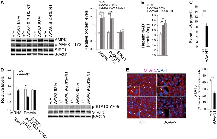
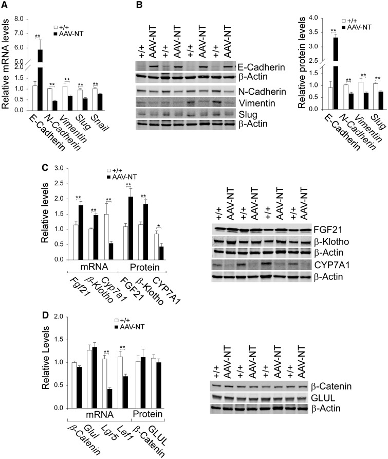
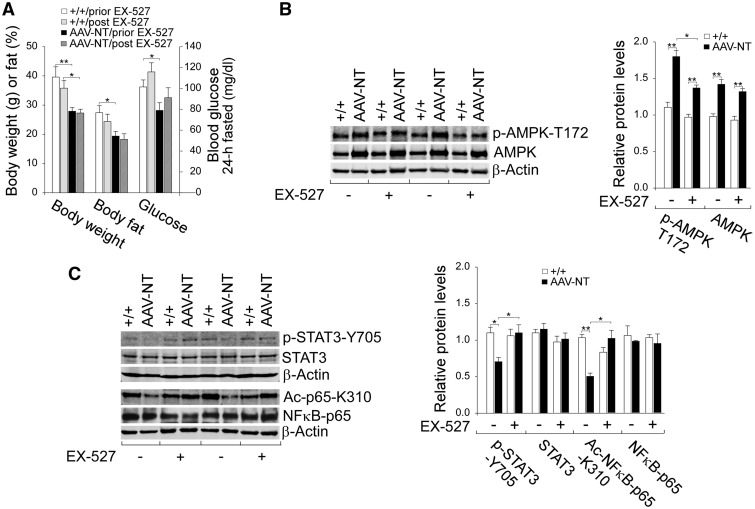
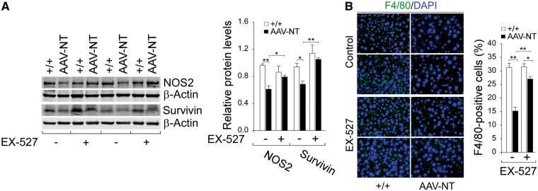
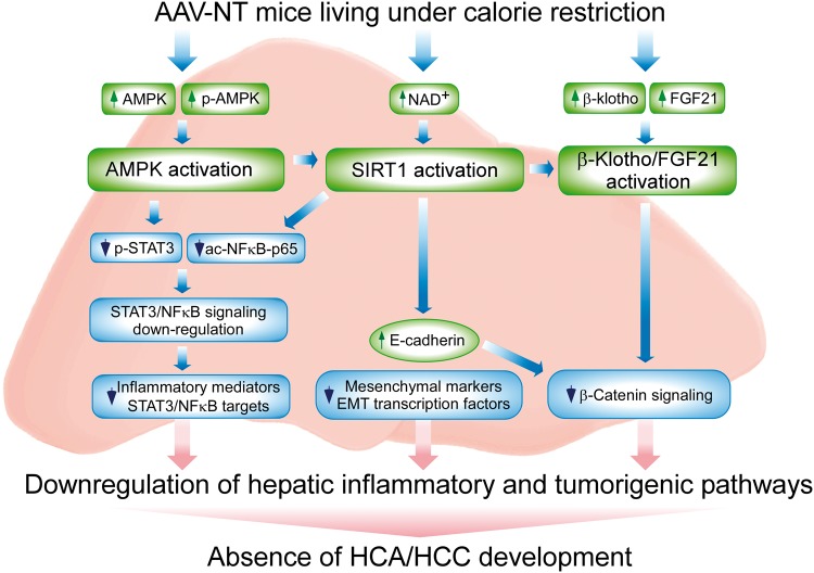
Glycogen storage disease type Ia (GSD-Ia) is characterized by impaired glucose homeostasis and long-term risks of hepatocellular adenoma (HCA) and carcinoma (HCC). We have shown that the non-tumor-bearing (NT), recombinant adeno-associated virus (rAAV) vector-treated GSD-Ia mice (AAV-NT mice) expressing a wide range (0.9-63%) of normal hepatic glucose-6-phosphatase-α activity maintain glucose homeostasis and display physiologic features mimicking animals living under calorie restriction (CR). We now show that in AAV-NT mice, the signaling pathways of the CR mediators, AMP-activated protein kinase (AMPK) and sirtuin-1 are activated. AMPK/sirtuin-1 inhibit the activity of STAT3 (signal transducer and activator of transcription 3) and NFκB (nuclear factor κB), the pro-inflammatory and cancer-promoting transcription factors. Sirtuin-1 also inhibits cancer metastasis via increasing the expression of E-cadherin, a tumor suppressor, and decreasing the expression of mesenchymal markers. Consistently, in AAV-NT mice, hepatic levels of active STAT3 and NFκB-p65 were reduced as were expression of mesenchymal markers, STAT3 targets, NFκB targets and β-catenin targets, all of which were consistent with the promotion of tumorigenesis. AAV-NT mice also expressed increased levels of E-cadherin and fibroblast growth factor 21 (FGF21), targets of sirtuin-1, and β-klotho, which can acts as a tumor suppressor. Importantly, treating AAV-NT mice with a sirtuin-1 inhibitor markedly reversed many of the observed anti-inflammatory/anti-tumorigenic signaling pathways. In summary, activation of hepatic AMPK/sirtuin-1 and FGF21/β-klotho signaling pathways combined with down-regulation of STAT3/NFκB-mediated inflammatory and tumorigenic signaling pathways can explain the absence of hepatic tumors in AAV-NT mice.


Figures

Figure 1
Figure 1
Analysis of hepatic AMPK, SIRT1 and STAT3 signaling in 66–88 week-old wild-type and rAAV-treated G6pc-/- mice. For quantitative RT-PCR, the data were analyzed for wild-type (+/+, n = 13) and AAV-NT (n = 22) mice. (A) Western-blot analysis of AMPK, p-AMPK-T172, SIRT1 and β-actin with quantification of protein levels by densitometry in wild-type (n = 8), AAV/3–63% (n = 4) and AAV/0.9–2.4%-NT (n = 4) mice. (B) Hepatic NAD+ levels in wild-type (n = 13), AAV/3–63% (N = 8), and AAV/0.9–2.4%-NT (n = 8) mice. (C) Blood IL-6 levels in wild-type (n = 13) and AAV-NT (n = 16) mice. (D) Quantification of Stat3 mRNA by real-time RT-PCR; Western blot analysis of p-STAT3-Y705, STAT3 and β-actin with quantification of protein levels by densitometry from 4 pairs of wild-type/AAV-NT mice. The AAV-NT analyzed included AAV/3–63% (n = 2) and AAV/0.9–2.4%-NT (n = 2) mice. (E) Immunofluorescence analysis of hepatic STAT3 nuclear localization and quantification of nuclear STAT3-translocated cells. Representative plates shown are analyzed for wild-type mice (n = 6) and AAV-NT (n = 5) mice. Scale bar: 25 μm. Arrows denote STAT3-positive nuclei. Data represent the mean ± SEM; **P < 0.005.
Figure 2
Figure 2
Analysis of hepatic NFκB signaling and inflammatory markers in 66–88 week-old wild-type and AAV-NT mice. For quantitative RT-PCR, the data were analyzed for wild-type (+/+, n = 13) and AAV-NT (n = 22) mice. For densitometry analysis, the data were analyzed from 4 pairs of wild-type/AAV-NT mice. The AAV-NT analyzed included AAV/3–63% (n = 2) and AAV/0.9–2.4%-NT (n = 2) mice. For blood MCP-1 and CRP levels, the data were analyzed for wild-type (+/+, n = 12) and AAV-NT (n = 22) mice. (A) Quantification Nfκb-p65 mRNA by real-time RT-PCR; Western blot analysis of NFκB-p65, Ac-NFκB-p65-K310 and β-actin with quantification of protein levels by densitometry. (B) Quantification of mRNA for STAT3/NFκB targets by real-time RT-PCR; Western-blot analysis of NOS2, survivin and β-actin with quantification of protein levels by densitometry. (C) Blood MCP-1 levels. (D) Immunofluorescence analysis of hepatic cells stained positive for F4/80 that detects macrophages and quantification of F4/80-positive cells. Representative plates shown are analyzed for wild-type (n = 6) and AAV-NT (n = 5) mice. Scale bar: 25 μm. (E) Quantification of Crp and Saa2 mRNA by real-time RT-PCR and blood CRP levels. Data represent the mean ± SEM; *P < 0.05; **P < 0.005.
Figure 3
Figure 3
Analysis of hepatic E-cadherin, mesenchymal markers, FGF21, β-klotho and β-catenin targets in 66–88 week-old wild-type and AAV-NT mice. For quantitative RT-PCR, the data were analyzed for wild-type (+/+, n = 13) and AAV-NT (n = 22) mice. For densitometry analysis, the data were analyzed from 4 pairs of wild-type/AAV-NT mice. The AAV-NT analyzed included AAV/3-63% (n = 2) and AAV/0.9–2.4%-NT (n = 2) mice. (A) Quantification of E-cadherin, N-cadherin, vimentin, Slug and Snail mRNA by real-time RT-PCR. (B) Western blot analysis of E-cadherin, N-cadherin, vimentin, Slug and β-actin with quantification of protein levels by densitometry. (C) Quantification β-Klotho, Fgf21 and Cyp7a1 mRNA by real-time RT-PCR; Western blot analysis of FGF21, β-klotho, CYP7A1 and β-actin; and quantification of protein levels by densitometry. (D) Quantification of β-Catenin, Glul, Lgr5, Cyclin D1 and Lef1 mRNA by real-time RT-PCR; Western-blot analysis of β-catenin, GLUL and β-actin; and quantification protein levels by densitometry. Data represent the mean ± SEM; *P < 0.05; **P < 0.005.
Figure 4
Figure 4
Effects of EX-527 on AMPK/STAT3/NFκB signaling in wild-type and AAV-NT mice. Wild-type and AAV-NT mice at age 64–74 weeks were treated daily for four weeks with EX-527 and their phenotype analyzed at age 68–78 weeks. For quantitative RT-PCR, the data were analyzed for wild-type (+/+, n = 8), wild-type-EX-527 (+/+/EX-527, n = 8), AAV-NT (n = 8) and AAV-NT-EX-527 (n = 8) mice. For densitometry analysis, the data were analyzed from 4 pairs of wild-type/AAV-NT and wild-type-EX-527/AAV-NT-EX-527 mice. (A) Body weight, body fat and fasting blood glucose levels of mice before and after EX-527 treatment. (B) Western blot analysis of p-AMPK-T172, AMPK and β-actin with quantification of protein levels by densitometry. (C) Western-blot analysis of p-STAT3-Y705, STAT3, NFκB-p65, Ac-NFκB-p65-K310 and β-actin and quantification protein levels by densitometry. Data represent the mean ± SEM; *P < 0.05; **P < 0.005.
Figure 5
Figure 5
Effects of EX-527 on hepatic STAT3/NFκB targets in wild-type and AAV-NT mice. Wild-type and AAV-NT mice at age 64–74 weeks were treated with EX-527 for 4 weeks, and their phenotype analyzed at age 68–78 weeks. For densitometry analysis, the data were analyzed from 4 pairs of wild-type/AAV-NT and wild-type-EX-527/AAV-NT-EX-527 mice. (A) Western blot analysis of NOS2, survivin, and β-actin with quantification of protein levels by densitometry. (B) Immunofluorescence analysis of hepatic cells stained positive for F4/80 that detects macrophages and quantification of F4/80-positive cells. Representative plates shown are analyzed for 4 pairs of wild-type/AAV-NT and wild-type-EX-527/AAV-NT-EX-527 mice. Scale bar: 25 μm. Data represent the mean ± SEM; *P < 0.05; **P < 0.005.
Figure 6
Figure 6
Effects of EX-527 on hepatic E-cadherin, mesenchymal markers, FGF21 and β-klotho in wild-type and AAV-NT mice. Wild-type and AAV-NT mice at age 64–74 weeks were treated with EX-527 for 4 weeks, and their phenotype analyzed at age 68–78 weeks. For densitometry analysis, the data were analyzed from 4 pairs of wild-type/AAV-NT and wild-type-EX-527/AAV-NT-EX-527 mice. (A) Western blot analysis of E-cadherin, N-cadherin, vimentin, slug and β-actin with quantification of protein levels by densitometry. (B) Western-blot analysis of FGF21, β-klotho, β-catenin, GLUL and β-actin and quantification protein levels by densitometry. Data represent the mean ± SEM; *P < 0.05; **P < 0.005.
Figure 7
Figure 7
Hepatic signaling pathways underlying the absence of HCA/HCC in G6pc-/- mice receiving gene therapy and living under calorie restriction. Several signaling pathways implicated in inflammation and tumorigenesis are downregulated in the liver of AAV-NT mice, contributing to the absence of hepatic tumors. The increases in total AMPK and p-AMPK-T172 activate AMPK that suppresses phosphorylation and activation of STAT3 signaling. The increase in hepatic NAD+ levels activates SIRT1 that suppresses NFκB signaling via deacetylation of the p65 subunit of NFκB. The down-regulation of hepatic STAT3/NFκB signaling leads to reduced expression of inflammatory and tumorigenic STAT3/NFκB targets. SIRT1 activation also negatively regulates tumor metastasis by increasing the expression of E-cadherin and reducing the expression of mesenchymal markers and the EMT transcription factors. The increase in E-cadherin also inhibits β-catenin signaling that plays a key role in the pathogenesis of HCC. CR also activates FGF21/β-klotho signaling that decreases the expression of β-catenin targets and prevents hepatocarcinogenesis.

References

    1. Chou J.Y., Jun H.S., Mansfield B.C. (2010) Glycogen storage disease type I and G6Pase-β deficiency: etiology and therapy. Nat. Rev. Endocrinol., 6, 676–688. - PMC - PubMed
    1. Rake J.P., Visser G., Labrune P., Leonard J.V., Ullrich K., Smit G.P. (2002) Glycogen storage disease type I: diagnosis, management, clinical course and outcome. Results of the European Study on Glycogen Storage Disease Type I (ESGSD I). Eur. J. Pediatr., 161 (Suppl 1), S20–S34. - PubMed
    1. Greene H.L., Slonim A.E., O’Neill J.A. Jr, Burr I.M. (1976) Continuous nocturnal intragastric feeding for management of type 1 glycogen-storage disease. N. Engl. J. Med., 294, 423–425. - PubMed
    1. Chen Y.T., Cornblath M., Sidbury J.B. (1984) Cornstarch therapy in type I glycogen-storage disease. N. Engl. J. Med., 310, 171–175. - PubMed
    1. Franco L.M., Krishnamurthy V., Bali D., Weinstein D.A., Arn P., Clary B., Boney A., Sullivan J., Frush D.P., Chen Y.T.. et al. (2005) Hepatocellular carcinoma in glycogen storage disease type Ia: a case series. J. Inherit. Metab. Dis., 28, 153–162. - PubMed

Publication types

MeSH terms

Supplementary concepts