. 2017 May 5;45(8):4619-4631.
doi: 10.1093/nar/gkx178.

Aicardi-Goutières syndrome protein TREX1 suppresses L1 and maintains genome integrity through exonuclease-independent ORF1p depletion

Affiliations

Aicardi-Goutières syndrome protein TREX1 suppresses L1 and maintains genome integrity through exonuclease-independent ORF1p depletion

Peng Li et al. Nucleic Acids Res. .

Abstract

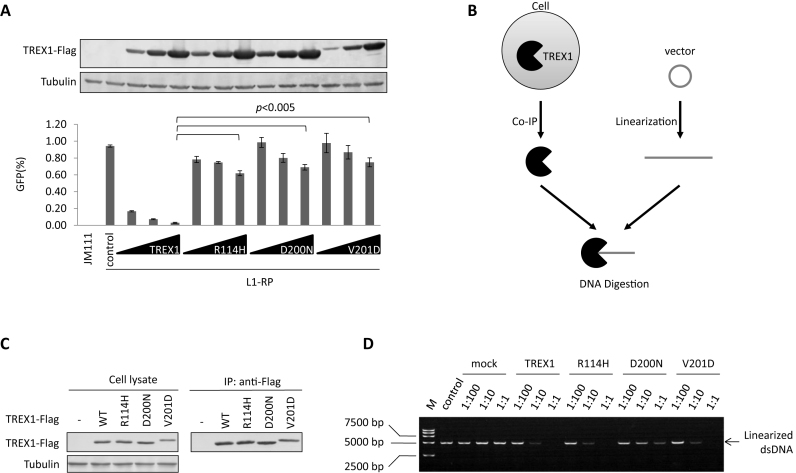
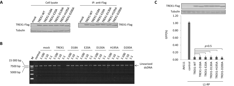
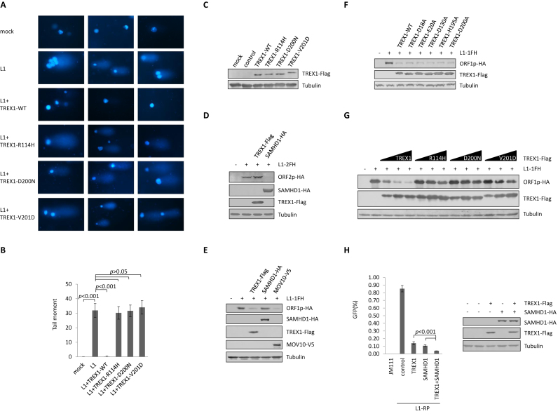
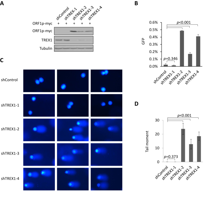
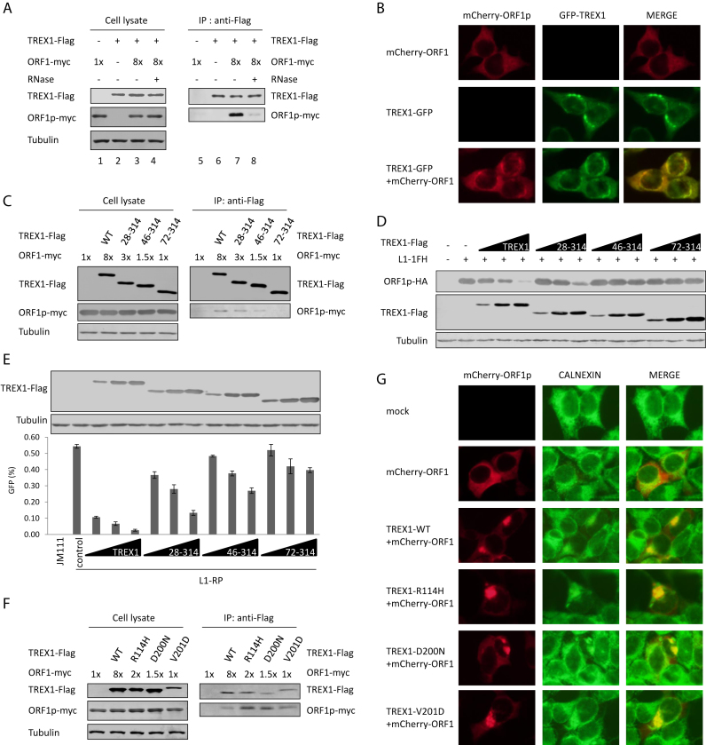
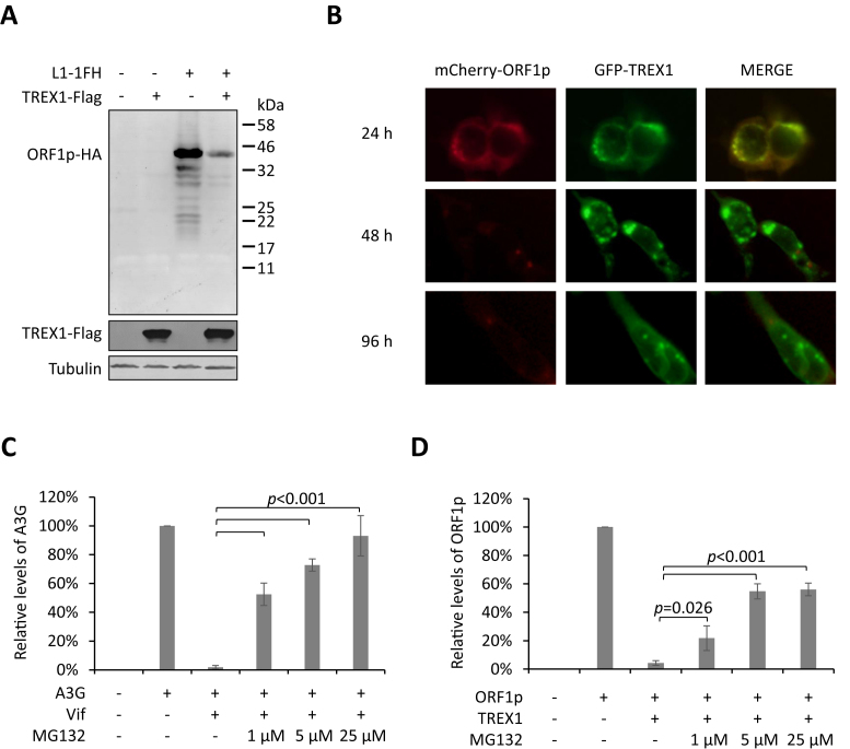
Maintaining genome integrity is important for cells and damaged DNA triggers autoimmunity. Previous studies have reported that Three-prime repair exonuclease 1(TREX1), an endogenous DNA exonuclease, prevents immune activation by depleting damaged DNA, thus preventing the development of certain autoimmune diseases. Consistently, mutations in TREX1 are linked with autoimmune diseases such as systemic lupus erythematosus, Aicardi-Goutières syndrome (AGS) and familial chilblain lupus. However, TREX1 mutants competent for DNA exonuclease activity are also linked to AGS. Here, we report a nuclease-independent involvement of TREX1 in preventing the L1 retrotransposon-induced DNA damage response. TREX1 interacted with ORF1p and altered its intracellular localization. Furthermore, TREX1 triggered ORF1p depletion and reduced the L1-mediated nicking of genomic DNA. TREX1 mutants related to AGS were deficient in inducing ORF1p depletion and could not prevent L1-mediated DNA damage. Therefore, our findings not only reveal a new mechanism for TREX1-mediated L1 suppression and uncover a new function for TREX1 in protein destabilization, but they also suggest a novel mechanism for TREX1-mediated suppression of innate immune activation through maintaining genome integrity.


Figures

Figure 1.
Figure 1.
AGS-associated exonuclease-active mutants of TREX1 do not suppress L1. (A) L1 assay results indicating that AGS-associated mutations significantly compromise TREX1's potency against L1 activity. Vectors expressing wild-type TREX1 or its AGS mutants in a dose manner (50, 150 and 450 ng) were co-transfected with 2 μg L1-RP into HEK293T cells seeded on a 12-well plate to examine the possible potency against L1 retrotransposition. At 4 days post-transfection, EGFP-positive cells were determined by flow cytometry. JM111 was used as the negative control for flow cytometry gating and VR1012 was the empty vector used as the negative control for TREX1 expression. The western blotting results (above) show the expressed levels of TREX1 and its mutants. (B) Cartoon showing the analysis of TREX1-mediated DNA digestion. FLAG-tagged TREX1 was expressed in HEK293T cells and purified through affinity chromatography. The target vector was first linearized then incubated with purified TREX1 protein for 20 min. The mixture was subjected to agarose electrophoresis for DNA detection. (C) Western blotting results showing the protein levels of TREX1 in both cell lysates and eluates in co-immunoprecipitation (co-IP). Three micrograms of vectors expressing TREX1 or its AGS mutants were transfected into HEK293T cells seeded on a 6-cm dish. At 48 h post-transfection, the cells were harvested for the IP assay shown in C and the DNase activity assay shown in D. (D) Agarose electrophoresis results showing that AGS-associated mutants R114H and V201D, but not D200N, can efficiently digest linearized DNA. M, DL15 000 DNA marker (Takara). Vector VR1012 was linearized and used as the plasmid substrate. The ratios shown are dilution rates of extract TREX1 protein. All the data shown in this figure are representative of at least three independent experiments. The error bars shown in A indicate the SD of three replicates within one experiment.
Figure 2.
Figure 2.
An exonuclease-independent mechanism contributes to TREX1-mediated L1 suppression. (A) Western blotting results showing the protein levels of TREX1 in both cell lysates and co-IP eluates. Three micrograms of vectors expressing TREX1 or its DNase-defective mutants were transfected into HEK293T cells seeded on a 6-cm dish. At 48 h post-transfection, the cells were harvested for the IP assay shown in A and DNase activity assay shown in B. (B) Agarose electrophoresis results showing that wild-type TREX1, and not its exonuclease-deactivated mutants, can digest linearized DNA. M, DL15 000 DNA marker (Takara). Linearized L1-RP was used in place of VR1012 as the substrate to confirm that the tested mutants did not affect the L1 assay results by compromising exonuclease activity. The ratios shown are dilution rates of extract TREX1 proteins. (C) L1 assay results showing that exonuclease-deactivated TREX1 mutants maintain potency against L1 replication. A total of 450 ng of VR1012 empty vector or vectors expressing TREX1 or its DNase-defective mutants were co-transfected with 2 μg L1-RP into HEK293T cells seeded on a 12-well plate to examine potency against L1 retrotransposition. At 4 days post-transfection, EGFP-positive cells were determined by flow cytometry. JM111 was used as the negative control for flow cytometry gating, and VR1012 was the empty vector used as the negative control for TREX1 expression. The western blotting results show the expressed levels of TREX1 and its mutants. All the data shown in this figure are representative of at least three independent experiments. The error bars shown in C indicate the SD of three replicates within one experiment.
Figure 3.
Figure 3.
TREX1 suppresses L1 activity and L1-induced genome nicking by reducing L1 ORF1p levels. (A) Comet assay results concerning TREX1 suppression of L1-induced genome damage. HeLa cells were transfected with L1-1FH (2 μg), along with 500 ng control vector or the vector expressing TREX1 or one of its mutants on a 12-well plate. The cells were then subjected to the comet assay and fluorescent imaging at 96 h post-transfection. The data are representative of three independent experiments and three random areas are shown for each sample from the same experiment. (B) The tail moment of the comets in A was analyzed for 100 cells for each sample using Comet Assay IV software. (C) Western blotting results indicating corresponding levels of TREX1 in each sample shown in A. (D) Western blotting results showing that TREX1 does not reduce the protein levels of ORF2p. TREX1 (500 ng) or SAMHD1 (500 ng) expression plasmids were co-transfected with L1-2FH (2 μg) into HEK293T cells seeded on a 12-well plate. At 48 h post transfection, the cells were harvested for western blotting. SAMHD1 was previously reported to reduce ORF2p and is shown as a positive control. (E) Western blotting results showing that TREX1 potently reduces protein levels of ORF1p. TREX1 (500 ng) or SAMHD1 (500 ng) or MOV10 (500 ng) expression plasmids were co-transfected with L1-1FH (300 ng) into HEK293T cells seeded on a 12-well plate. At 48 h post -transfection, the cells were harvested for western blotting. SAMHD1 was introduced as a negative control, and MOV10 as a positive control (30,59). (F) Western blotting results showing that exonuclease-deactivated TREX1 mutants maintain the ability to reduce ORF1p levels. Five hundred nanogram TREX1 WT or DNase mutant expression plasmids were co-transfected with L1-1FH (300 ng) into HEK293T cells seeded on a 12-well plate. At 48 h post transfection, the cells were harvested for western blotting. (G) Western blotting results showing that AGS-associated mutations compromise TREX1's ability to deplete ORF1p. Vectors expressing TREX1 or its AGS mutants in a dose manner (50, 150 and 450 ng) were co-transfected with L1-1FH (300 ng) into HEK293T cells seeded on a 12-well plate. At 48 h post transfection, the cells were harvested for western blotting. (H) L1 assay results indicating that the suppressive effects of TREX1 and SAMHD1 against L1 replication are additive. Vectors expressing TREX1 (500 ng) or SAMHD1 (500 ng) or both plasmids were co-transfected with 2 μg L1-RP into HEK293T cells seeded on a 12-well plate to examine possible potency against L1 retrotransposition. The western blotting results show the expressed levels of TREX1 and SAMHD1. All the data shown in this figure are representative of at least three independent experiments. Three random areas are shown for each sample from the same experiment in A. The error bars shown in B indicate the standard error of the mean (SEM) of three independent experiments, and those in H indicate the SD of three replicates within one experiment.
Figure 4.
Figure 4.
Endogenous TREX1 destabilizes ORF1p, suppresses L1 activity and protects genomic integrity. (A) Western blotting results showing that the levels of ORF1p protein are increased in HeLa cells in which endogenous TREX1 has been knocked down. ORF1p expression plasmid (300 ng) was transfected into five HeLa cell lines (HeLa shControl and four HeLa shTREX1 cells) seeded on a 12-well plate. At 48 h post-transfection, the cells were harvested for western blotting. (B) L1 assay results indicating that reducing endogenous TREX1 levels in HeLa cells results in an increase in L1-RP activity. Two micrograms of L1–RP were transfected into HeLa cells with shControl or shTREX1 constructs to examine the possible potency of L1 retrotransposition. (C) Results from comet assays suggesting that endogenous TREX1 protects genomic integrity. (D) The tail moment of the comets in C were analyzed for 100 cells for each sample using Comet assay IV software. All the data shown in this figure are representative of three independent experiments. Three random areas are shown for each sample from the same experiment in C. The error bars shown in B indicate the S\D of three replicates within one experiment, and those in D indicate the SEM of three independent experiments.
Figure 5.
Figure 5.
The TREX1–ORF1p interaction is important but not sufficient for TREX1-mediated depletion of ORF1p. (A) Co-IP results indicating that TREX1 interacts with ORF1p in an RNA-dependent manner. ORF1p (600 ng was 1×) and/or TREX1 (2 μg) expression plasmids were transfected into HEK293T cells seeded on 6-cm dishes. The transfection dose of the ORF1p-expressing vector was 8-fold higher than that for TREX1 (lanes 3 and 4) in order to achieve similar protein expression levels (lane 2). (B) Fluorescence imaging results showing the co-localization of TREX1 (green) and ORF1p (red) in live cells. TREX1-GFP (1 μg) and/or mCherry-ORF1 (1 μg) expression plasmids were transfected into HEK293T cells seeded on a 6-well plate. (C) Co-IP results showing that N-terminal truncations compromise TREX1's ability to interact with ORF1p. Two micrograms of vectors expressing TREX1 or its N-terminal truncations were co-transfected with the indicated amount of ORF1p (600 ng was 1×) expression plasmids into HEK293T cells seeded on 6-cm dishes. (D) Western blotting results indicating that N-terminal truncations compromise TREX1's ability to deplete ORF1p. Vectors expressing TREX1 or its N-terminal truncations in different doses (50, 150 and 450 ng) were co-transfected with L1-1FH (300 ng) into HEK293T cells seeded on a 12-well plate. At 48 h post transfection, the cells were harvested for western blotting. (E) L1 assay results indicating that N-terminal truncations compromise TREX1's ability to suppress L1. Vectors expressing TREX1 or its N-terminal truncations in different doses (50, 150, and 450 ng) were co-transfected with 2 μg L1-RP into HEK293T cells seeded on a 12-well plate to examine the possible potency of L1 retrotransposition. At 4 days post-transfection, EGFP-positive cells were determined by flow cytometry. JM111 was used as the negative control for flow cytometry gating and VR1012 was the empty vector used as the negative control for TREX1 expression. The western blotting results show the expressed levels of TREX1 and its mutants. (F) Co-IP results showing that AGS-associated TREX1 mutants maintain the ability to interact with ORF1p. Various doses of the ORF1p expression vector were used to achieve similar expression in the presence of different TREX1 mutants. Two micrograms of vectors expressing TREX1 or its AGS mutants were co-transfected with the indicated amount of ORF1p (600 ng was 1×) expression plasmids into HEK293T cells seeded on 6-cm dishes. (G) Fluorescence imaging results show that AGS-associated mutants still alter the subcellular localization of ORF1p (red); calnexin (green) is a protein marker labeling the endoplasmic reticulum (ER) (8). One microgram of VR1012 vector or vectors expressing TREX1 or its AGS mutants were co-transfected with 1 μg mCherry-ORF1 expression plasmid into HEK293T cells seeded on a 6-well plate. All the data shown in this figure are representative of at least three independent experiments. The error bars shown in E indicate the SD of three replicates within one experiment.
Figure 6.
Figure 6.
Proteasome proteolysis is involved in TREX1-mediated removal of ORF1p. (A) Full-length western blot showing that no smaller fragments of ORF1p are increased in concentration in the presence of TREX1. TREX1 (500 ng) and/or L1-1FH (300 ng) expression plasmids were transfected into HEK293T cells seeded on a 12-well plate. (B) Fluorescence imaging results for live cells showing that TREX1-induced ORF1p depletion occurs after a 24 h delay. HEK293T cells were co-transfected with ORF1p-mCherry and TREX1-EGFP expression vectors and subjected to fluorescence imaging at 24, 48 and 96 h post-transfection. TREX1-GFP (1 μg) and/or mCherry-ORF1 (1 μg) expression plasmids were transfected into HEK293T cells seeded on a 6-well plate. (C) Bar graph showing that proteasome inhibitor MG132 potently rescues APOBEC3G from Vif-induced degradation. VR1012 (900 ng) empty vector or Vif (900 ng) expression plasmid was co-transfected with A3G (300 ng) into HEK293T cells seeded on a 12-well plate. (D) Bar chart indicating that proteasome inhibitor MG132 rescues ORF1p from TREX1-mediated depletion. VR1012 (500 ng) empty vector or TREX1 (500 ng) expression plasmid was co-transfected with L1-1FH (300 ng) into HEK293T cells seeded on a 12-well plate. Data shown in A and B are representative of at least three independent experiments. Protein levels shown in B and D were quantified with ImageJ software and normalized to the levels of tubulin (as the loading control). Mean (± SE) values from three independent tests are shown.

Similar articles

Cited by

References

    1. Gehrke N., Mertens C., Zillinger T., Wenzel J., Bald T., Zahn S., Tuting T., Hartmann G., Barchet W.. Oxidative damage of DNA confers resistance to cytosolic nuclease TREX1 degradation and potentiates STING-dependent immune sensing. Immunity. 2013; 39:482–495. - PubMed
    1. Hiller B., Achleitner M., Glage S., Naumann R., Behrendt R., Roers A.. Mammalian RNase H2 removes ribonucleotides from DNA to maintain genome integrity. J. Exp. Med. 2012; 209:1419–1426. - PMC - PubMed
    1. Lazzaro F., Novarina D., Amara F., Watt D.L., Stone J.E., Costanzo V., Burgers P.M., Kunkel T.A., Plevani P., Muzi-Falconi M.. RNase H and postreplication repair protect cells from ribonucleotides incorporated in DNA. Mol. Cell. 2012; 45:99–110. - PMC - PubMed
    1. Reijns M.A., Rabe B., Rigby R.E., Mill P., Astell K.R., Lettice L.A., Boyle S., Leitch A., Keighren M., Kilanowski F. et al. . Enzymatic removal of ribonucleotides from DNA is essential for mammalian genome integrity and development. Cell. 2012; 149:1008–1022. - PMC - PubMed
    1. Luan D.D., Korman M.H., Jakubczak J.L., Eickbush T.H.. Reverse transcription of R2Bm RNA is primed by a nick at the chromosomal target site: a mechanism for non-LTR retrotransposition. Cell. 1993; 72:595–605. - PubMed

Publication types

MeSH terms

Supplementary concepts

LinkOut - more resources