. 2017 Jun 2;45(10):6147-6167.
doi: 10.1093/nar/gkx168.

The primary transcriptome of Neisseria meningitidis and its interaction with the RNA chaperone Hfq

Affiliations

The primary transcriptome of Neisseria meningitidis and its interaction with the RNA chaperone Hfq

Nadja Heidrich et al. Nucleic Acids Res. .

Abstract

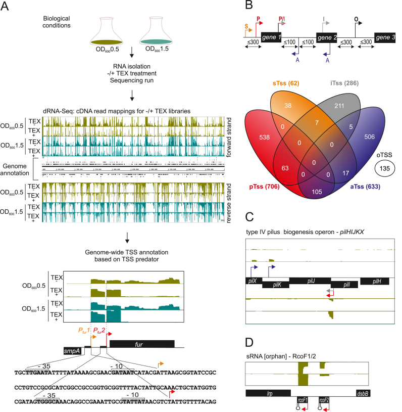
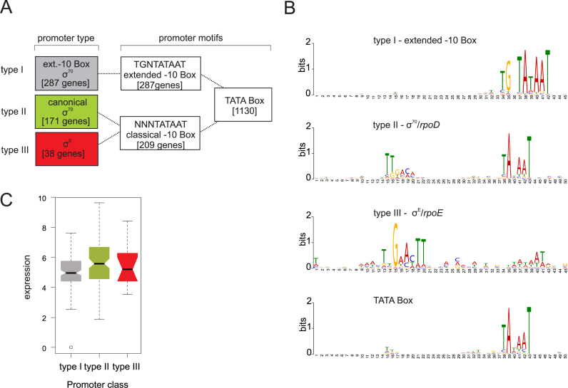
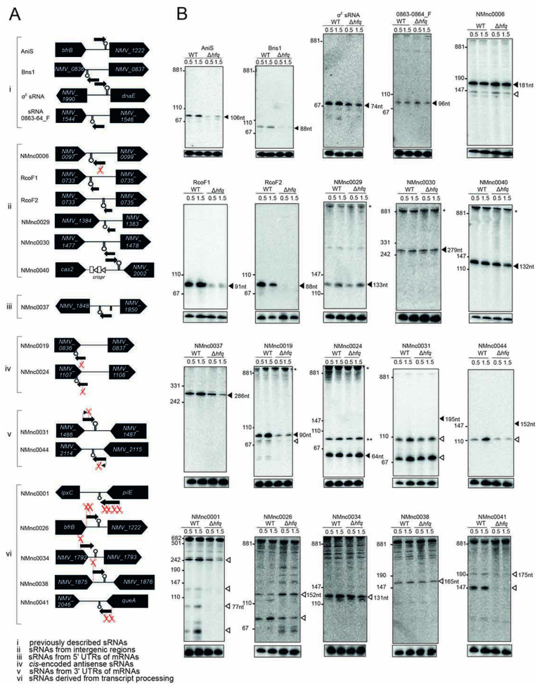
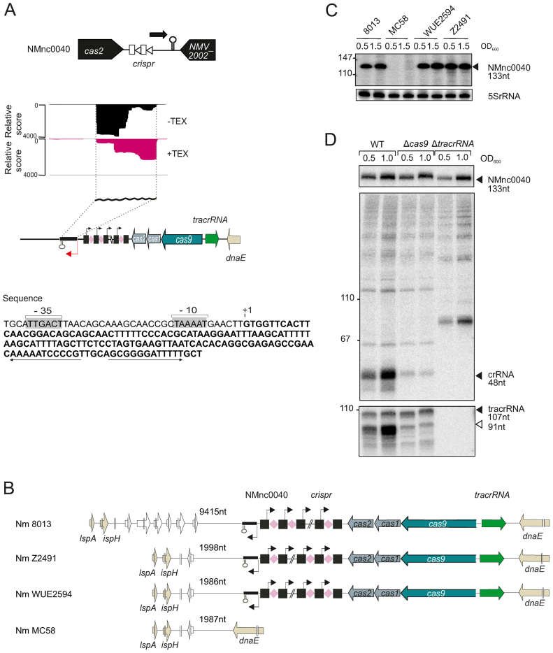
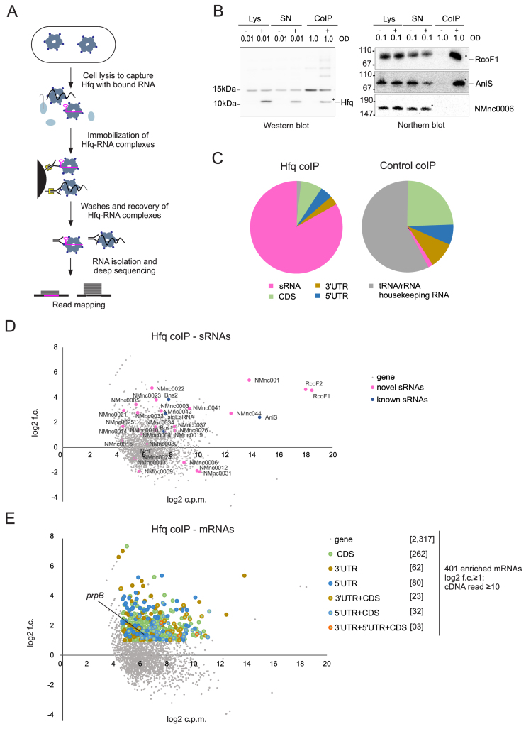
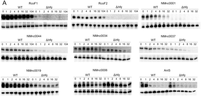
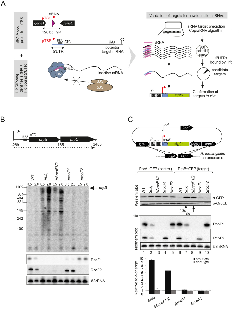
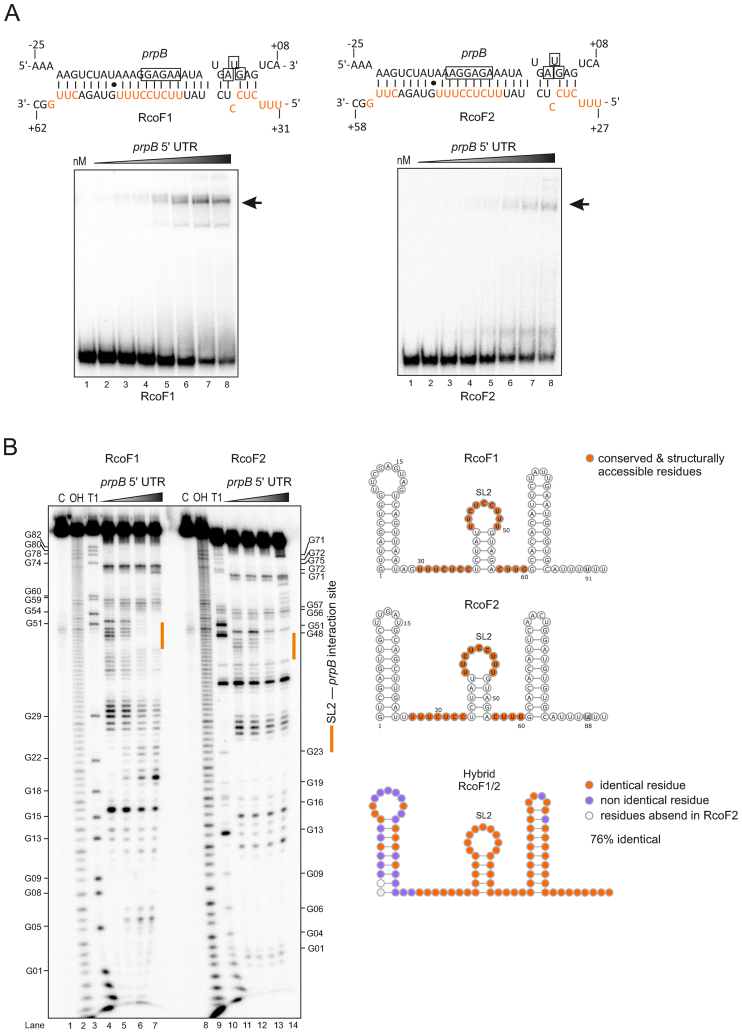
Neisseria meningitidis is a human commensal that can also cause life-threatening meningitis and septicemia. Despite growing evidence for RNA-based regulation in meningococci, their transcriptome structure and output of regulatory small RNAs (sRNAs) are incompletely understood. Using dRNA-seq, we have mapped at single-nucleotide resolution the primary transcriptome of N. meningitidis strain 8013. Annotation of 1625 transcriptional start sites defines transcription units for most protein-coding genes but also reveals a paucity of classical σ70-type promoters, suggesting the existence of activators that compensate for the lack of -35 consensus sequences in N. meningitidis. The transcriptome maps also reveal 65 candidate sRNAs, a third of which were validated by northern blot analysis. Immunoprecipitation with the RNA chaperone Hfq drafts an unexpectedly large post-transcriptional regulatory network in this organism, comprising 23 sRNAs and hundreds of potential mRNA targets. Based on this data, using a newly developed gfp reporter system we validate an Hfq-dependent mRNA repression of the putative colonization factor PrpB by the two trans-acting sRNAs RcoF1/2. Our genome-wide RNA compendium will allow for a better understanding of meningococcal transcriptome organization and riboregulation with implications for colonization of the human nasopharynx.


Figures

Figure 1.
Figure 1.
Differential RNA-seq and growth-phase based annotation of transcriptional start sites (TSSs). (A) (top) Schematic workflow for generating the biological, library and sequencing samples from strain N. meningitidis 8013 at two different growth phases (OD600 0.5 and 1.5) that were subjected to dRNA-seq analysis for genome-wide TSS annotation based on TSSpredator. (bottom) dRNA‐seq data from two growth phases OD600 0.5 and 1.5 were mapped to the fur gene. Red and orange arrows indicate a primary (pTSS) and secondary (sTSS) transcriptional start site and confirm initiation of RNA transcription as mapped with primer extension experiments in (42). Nucleotide sequence of the smpA- fur intergenic region (IGR) with promoter DNA elements are boxed and marked −10 and −35 at Pfur1 and Pfur2. (B) (Top) TSS classifications based on expression strength and genomic context: primary (P, red), secondary (S, orange), internal (I, gray), antisense (A, blue) or orphan (O, black). Venn diagrams were generated by VENNY (http://bioinfogp.cnb.csic.es/tools/venny/index.html). (C) Comparative annotation of primary TTSs (pTSSs) of the pilXKJIH operon. Detection of a pTSS (red arrow) for pilJ within the operon at two growth phases OD600 0.5 and 1.5 indicates a different genetic organization of their transcriptional units. Gray and blue arrows indicate an iTSS and an antisense TSS (aTSS), respectively. (D) Detection of an orphan TSS (oTSS; not associated with annotation) derived from dRNA-seq reads mapped to the intergenic lrp-dsbB region reveals two non-coding RNAs RcoF1 and RcoF2.
Figure 2.
Figure 2.
Promoter motifs detected for TSS of N. meningitidis 8013 and expression strength. (A) Schematic overview of promoter types derived from a motif search based on 1625 TSS upstream sequences of N. meningitidis 8013. The number of motifs for each type of promoter detected for the two sigma factors σD and σE in the upstream sequences of the TSS are indicated. (B) Promoter motifs for each type within a fixed size of either 45 or 50 nt, that was applied for the search using the MEME toolkit are shown. (C) Box-and-whiskers plot depicting differences in the expression (log (basemean)) between genes with the three different promoter types. The line within each box gives the median and the upper and lower margins the upper and the lower quartile, respectively. The whiskers denote the highest and the lowest values, respectively, and the open circles outliers.
Figure 3.
Figure 3.
Expression analysis of candidate sRNAs in N. meningitidis 8013. Total RNA was extracted at mid logarithmic (OD600 = 0.5) and late logarithmic/early-stationary (OD600 = 1.5) growth phases from wild-type (WT) and Δhfq N. meningitidis strains and analyzed by northern blot using labeled DNA probes complementary to the sRNAs (see Supplementary Table S3). (A) The genomic locations and relative orientations of (i) previously described sRNAs, (ii) sRNAs from IGR, (iii) sRNAs from 5΄ untranslated regions (UTRs) of mRNAs, (iv) cis-encoded antisense sRNAs, (v) sRNAs from 3΄ UTRs of mRNAs and (vi) sRNAs derived from transcript processing are shown. The designation in (vi) is the same as in (v) except that the promoters are shared between the sRNA and mRNA. Here, the mRNAs undergo ribonucleolytic cleavage to yield the sRNAs. Genes and sRNAs are shown in black. Arrows and scissors indicate TSS and processing sites, respectively. Black rectangle indicates the promoter of NMV_1848 and NMnc0037, respectively. (B) Gel images of blots from previously described sRNAs (i) and different classes of candidate sRNAs (ii–vi). The blots were probed for the housekeeping 5S rRNA as loading control. Filled triangles indicate bands for sRNAs derived from TSS and open triangles indicate bands derived from processing. Asteriks indicate signals of unclear origin, may represent longer read-through transcripts at the locus, result from cross hybridization of the probe with other abundant transcripts, or stem from undigested DNA trapped in the slot.
Figure 4.
Figure 4.
A strain-specific sRNA, NMnc0040 is linked to the CRISPR-Cas locus. (A) (top) Genomic location of NMnc0040 sRNA. dRNA–seq reads of −/+ TEX libraries mapped to NMnc0040 sRNA. (bottom) Nucleotide sequence of NMnc0040 is indicated in bold letters. The −10 and −35 sites of the NMnc0040 promoter are boxed in light gray. ‘+1’ marks the TSS and the transcribed NMnc0040 sequence is given in bold letters. The rho-independent terminator is indicated by arrows. (B) Conservation analysis of NMnc0040 in four prototypical N. meningitidis strains. NMnc0040 is absent in MC58. In the WEU2594, Z2491 and 8013 strains NMnc0040 is linked to CRISPR-Cas locus and located upstream of the CRISPR array. The flanking gene at the 3΄ end is variable. Distances to flanking genes are indicated in nucleotides. (C) Total RNA from the four N. meningitidis strains 8013, MC58, WUE2594 and Z2491 belonging to three different serogroups was isolated at mid logarithmic (OD600 = 0.5) and late logarithmic/early stationary growth phase (OD600 = 1.5) and analyzed by northern blot using labeled DNA probes complementary to NMnc0040 (see Supplementary Table S3). The blots were probed for the housekeeping 5S rRNA as loading control. (D) Northern blot analysis of NMnc0040 sRNA, tracrRNA and crRNAs in 8013 WT, Δcas9 and ΔtracrRNA strains. Total RNA (5 μg per lane) isolated from WT, ΔtracrRNA and Δcas9 at mid logarithmic (OD600 = 0.5) and late logarithmic/early stationary growth phase (OD600 = 1.5) was analyzed on a northern blot using labeled DNA probes complementary to NMnc0040, crRNA and tracrRNA in N. meningitidis 8013.
Figure 5.
Figure 5.
The repertoire of Hfq regulated sRNAs and mRNAs in N. meningitidis 8013. (A) Schematic workflow for generating co-immunoprecipitation (coIP) samples of WT and 3× FLAG tagged hfq strains grown in rich media to mid log phases (OD600 = 0.5) in two independent experiments. (B) Quality of RNA and protein samples was analyzed by western blots and northern blots. Lysate (Lys), supernatant (SN) and coIP samples were obtained from pull-down experiments with mouse anti-FLAG antibody performed in the presence (+) or absence (−) of the 3× FLAG tagged Hfq protein. OD600 equivalents of protein and RNA samples loaded on the gel are shown. Western blot with rabbit anti-FLAG antibody (left panels) and northern blot with probe against RcoF1 and AniS sRNAs as well as NMnc0006 sRNA as negative control (right panel) confirmed the success of Hfq pull-downs. The band indicated by a star within the coIP (+) lanes correspond to the sizes of purified Hfq proteins and co-purified RcoF1 sRNA. Size markers are given on the left (in kDa). (C) Pie chart for Hfq coIP and control coIP showing the relative proportions of all Hfq-associated sequences that unequivocally mapped to different classes of RNA sequences. (D) Scatter-plot of RIP-seq results. Axes represent log fold-change between the control coIP and Hfq coIP (y-axis) and abundance in log counts per million (x-axis) of cDNA reads obtained. New sRNAs and previously described sRNAs are depicted by pink and blue dots, respectively. (E) Scatter-plot analysis of RIP-seq results depicting 401 mRNAs and their associated segments (CDS, 5΄ UTR and 3΄ UTR) that were enriched (log2 f.c. ≥1; cDNA read ≥10) in the Hfq coIP, for example the 5΄ UTR of prpB gene. Axes are identical to above.
Figure 6.
Figure 6.
Stability of Hfq binding sRNAs. (A) sRNA half-lives for RcoF1, RcoF2, NMnc0001, NMnc0044, NMnc0034, NMnc0037, NMnc0019, NMnc006 and AniS. At the top of each panel, northern blots of total RNA extracted at the indicated time points (minutes) after addition of rifampicin (250 μg/ ml) to terminate transcription in a N. meningitidis 8013 WT and an isogenic Δhfq strain is shown. Prior to the addition of rifampicin, strains were grown to OD600 of 0.5. 5S rRNA served as loading control.
Figure 7.
Figure 7.
In vivo validation of prpB regulation by the two sRNAs RcoF1 and RcoF2. (A) (Left) Schematic representation for combining data gained from dRNA-seq and Hfq RIP-seq analysis to uncover and validate targets for newly identified sRNAs. dRNA-seq enriches for primary transcriptional start sides (pTSS) as indicated by red arrows and therewith uncovers sRNAs (pink triangles) located in IGR and other transcriptome features like 5΄ UTRs of mRNAs colored in blue. Hfq RIP-seq allows for the identification of sRNAs and 5΄ UTRs as targets of Hfq. 5΄ UTRs are target sites for Hfq-dependent sRNA regulation that leads to inhibition of ribosome binding, resulting in reduced translation. (Right) Schematic workflow for target prediction by the copra algorithm and validation via a superfolder gfp (sfgfp) reporter gene fusion, exemplified by the two newly identified sRNAs RcoF1 and RcoF2 and their target prpB. Hfq-bound 5΄ UTRs of mRNAs identified by Hfq-RIPseq (colored in blue) serve to further screen potential mRNA targets predicted for new sRNAs identified by dRNA-seq (colored in pink). (B) (top) The 1165-nt-long prpB transcript is part of the dicistronic mRNA prpB-prpC (dotted line) encoding a methylcitrate lyase and methylcitrate synthase, respectively. The TSS is indicated by arrows and the ribosome binding site (RBS) by a black bar. Numbers indicate the distance to the TSS. (bottom) The expression of prpB was analyzed during two growth phases in WT, Δhfq, ΔΔrcof1/2 double deletion mutants as well as Δrcof1 and Δrcof2 single deletion mutants at indicated optical densities (OD600) using a 32P-UTP labeled riboprobe. RcoF1 and RcoF2 were detected with JVO13282 and JVO13283. 5S rRNA served as loading control. (C) (top) Schematic illustration of the integrational vector pGCC2 for construction of translational superfolder gfp (sfgfp) fusions that were inserted into the lctP and aspC locus of N. meningitidis 8013 (127). The prpB 5΄ UTR and the first 15 amino acids of the prpB coding region (gray box) were fused to sfgfp reporter gene (green box) that is transcribed from the constitutive PLtetO-1 promoter as indicated. Individual elements are not drawn to scale. A sfgfp reporter fusion expressing the 5΄ UTR and the first 15 amino acids of the porA gene served as control. (bottom) N. meningitidis 8013 WT, Δhfq, ΔΔrcof1/2 double deletion mutants as well as Δrcof1 and Δrcof2 single deletion mutants expressing either the target-gfp or the control-gfp fusions were grown to mid logarithmic phase and RNA and protein samples were analyzed by northern blot and western blot. RcoF1 was detected with JVO-13282 and RcoF2 was detected with JVO-13283. 5S rRNA served as loading control. Whole cell protein fractions (OD600 = 0.01 for western blot) were detected with mouse anti-Gfp antiserum. GroEL served as control. Relative fold expression changes of prpB::gfp and porA::gfp (control) fusions in Δhfq, of both sRNAs RcoF1 and RcoF2 or of each sRNA alone determined by western blot analysis for Gfp in comparison with the respective WT backgrounds are represented in the bar plot. Error bars indicate the SDs among three biological replicates.
Figure 8.
Figure 8.
In vitro gel-shift and structure probing assay show a direct interaction between the two sRNAs RcoF1/2 and the prpB leader. (A) (top) Predicted base-pairing interactions of RcoF1 and RcoF2 with their respective target mRNA prpB using RNA hybrid (79). The Shine-Dalgarno sequences and start codons of the mRNAs are boxed. Conserved and structurally accessible residues are shown in red. The proposed strength of interaction for each RNA duplex is −36.6 kcal/mol. (bottom) Gels-shift experiment with in vitro synthesized sRNAs (RcoF1/2) and prpB mRNA leader (−289 to +42 relative to the annotated start codon). About 0.04 pmol of each 32P-labeled sRNA was incubated with increasing concentrations of unlabeled prpB mRNA leader (lane 1–8: 0, 8, 16, 62.5, 125, 250, 500, 1000 nM). Arrows indicate the RNA-RNA duplex (B) (Left) In-line probing of 0.04 pmol of each 32P-labeled sRNA in the absence (lane 4, lane 11) or presence (lane 5–7, lane 12–14) of prpB mRNA leader. Untreated RNA (lane C), partially alkali (lane OH) or RNase T1 (lane T1) digested RcoF1 and RcoF2 served as ladders. (Right) Predicted secondary structures of sRNAs RcoF1 and RcoF2 using RNAfold. For both sRNAs nucleotides located within three single stranded regions (marked in orange) were predicted to base-pair with the 5΄ UTR of a dicistronic prpB-prpC mRNA. Identical residues (marked in orange) and non identical residues (marked in purple) of both sRNAs are represented by the hybrid sRNA structure. The designation SL2 represents loop 2 of the second Stem-loop structure.

Similar articles

Cited by

References

    1. Henriques-Normark B., Normark S.. Commensal pathogens, with a focus on Streptococcus pneumoniae, and interactions with the human host. Exp. Cell Res. 2010; 316:1408–1414. - PubMed
    1. Stephens D.S., Greenwood B., Brandtzaeg P.. Epidemic meningitis, meningococcaemia, and Neisseria meningitidis. Lancet. 2007; 369:2196–2210. - PubMed
    1. Caugant D.A., Maiden M.C.J.. Meningococcal carriage and disease-population biology and evolution. Vaccine. 2009; 27:B64–B70. - PMC - PubMed
    1. Greenwood B. Manson lecture. Meningococcal meningitis in africa. Trans. R. Soc. Trop. Med. Hyg. 1999; 93:341–353. - PubMed
    1. Fredricks D.N., Relman D.a.. Sequence-based identification of microbial pathogens: a reconsideration of Koch's postulates. Clin. Microbiol. Rev. 1996; 9:18–33. - PMC - PubMed

MeSH terms