

Clinical and genetic characterization of leukoencephalopathies in adults
- PMID: 28334938
- PMCID: PMC5405235
- DOI: 10.1093/brain/awx045
Clinical and genetic characterization of leukoencephalopathies in adults
Abstract
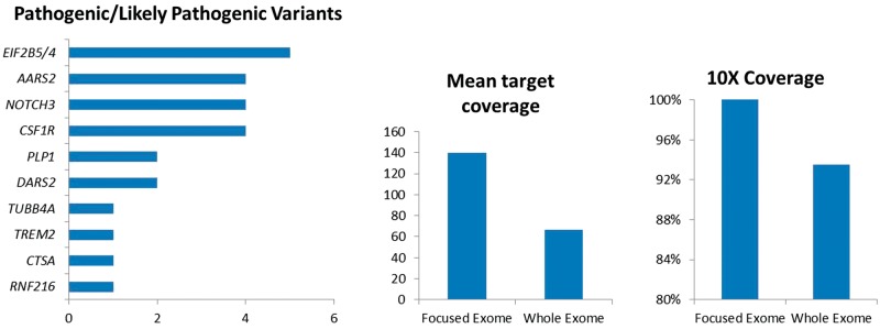
Leukodystrophies and genetic leukoencephalopathies are a rare group of disorders leading to progressive degeneration of cerebral white matter. They are associated with a spectrum of clinical phenotypes dominated by dementia, psychiatric changes, movement disorders and upper motor neuron signs. Mutations in at least 60 genes can lead to leukoencephalopathy with often overlapping clinical and radiological presentations. For these reasons, patients with genetic leukoencephalopathies often endure a long diagnostic odyssey before receiving a definitive diagnosis or may receive no diagnosis at all. In this study, we used focused and whole exome sequencing to evaluate a cohort of undiagnosed adult patients referred to a specialist leukoencephalopathy service. In total, 100 patients were evaluated using focused exome sequencing of 6100 genes. We detected pathogenic or likely pathogenic variants in 26 cases. The most frequently mutated genes were NOTCH3, EIF2B5, AARS2 and CSF1R. We then carried out whole exome sequencing on the remaining negative cases including four family trios, but could not identify any further potentially disease-causing mutations, confirming the equivalence of focused and whole exome sequencing in the diagnosis of genetic leukoencephalopathies. Here we provide an overview of the clinical and genetic features of these disorders in adults.
Keywords: imaging; leukodystrophy; neurodegeneration; white matter lesion.
© The Author (2017). Published by Oxford University Press on behalf of the Guarantors of Brain.
Figures
References
-
- Ahmed RM, Murphy E, Davagnanam I, Parton M, Schott JM, Mummery CJ et al. A practical approach to diagnosing adult onset leukodystrophies. J Neurol Neurosurg Psychiatry 2014; 85: 770–81. - PubMed
-
- Bugiani M, Kevelam SH, Bakels HS, Waisfisz Q, Ceuterick-de Groote C, Niessen HW et al. Cathepsin A-related arteriopathy with strokes and leukoencephalopathy (CARASAL). Neurology 2016; 87: 1777–86. - PubMed
-
- Hurst S, Garbern J, Trepanier A, Gow A. Quantifying the carrier female phenotype in Pelizaeus-Merzbacher disease. Genet Med 2006; 8: 371–8. - PubMed
-
- Lynch DS, Zhang WJ, Lakshmanan R, Kinsella JA, Uzun GA, Karbay M et al. Analysis of mutations in AARS2 in a series of CSF1R -negative patients with adult-onset leukoencephalopathy with axonal spheroids and pigmented glia. JAMA Neurol 2016; 114: 494–500. - PubMed
MeSH terms
Grants and funding
LinkOut - more resources
Full Text Sources
Other Literature Sources
Molecular Biology Databases
Miscellaneous
