. 2017 Apr 15;26(8):1522-1534.
doi: 10.1093/hmg/ddx057.

DNMT1 mutations found in HSANIE patients affect interaction with UHRF1 and neuronal differentiation

DNMT1 mutations found in HSANIE patients affect interaction with UHRF1 and neuronal differentiation

Martha Smets et al. Hum Mol Genet. .

Abstract

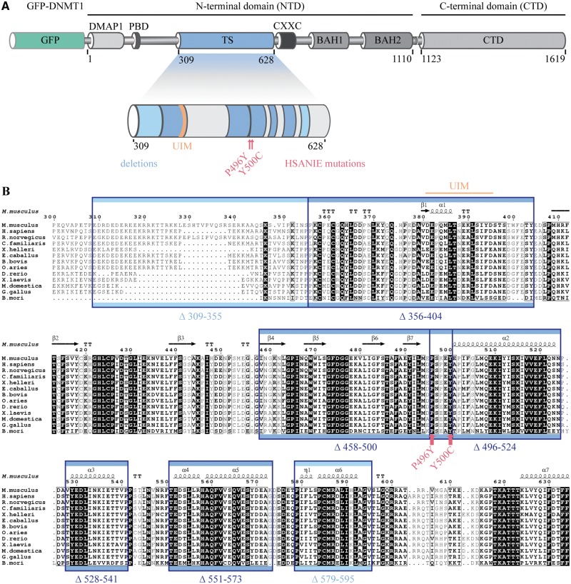
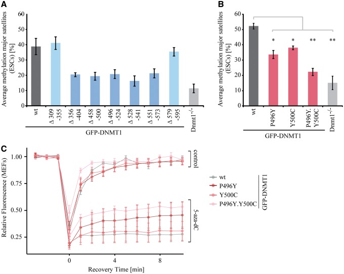
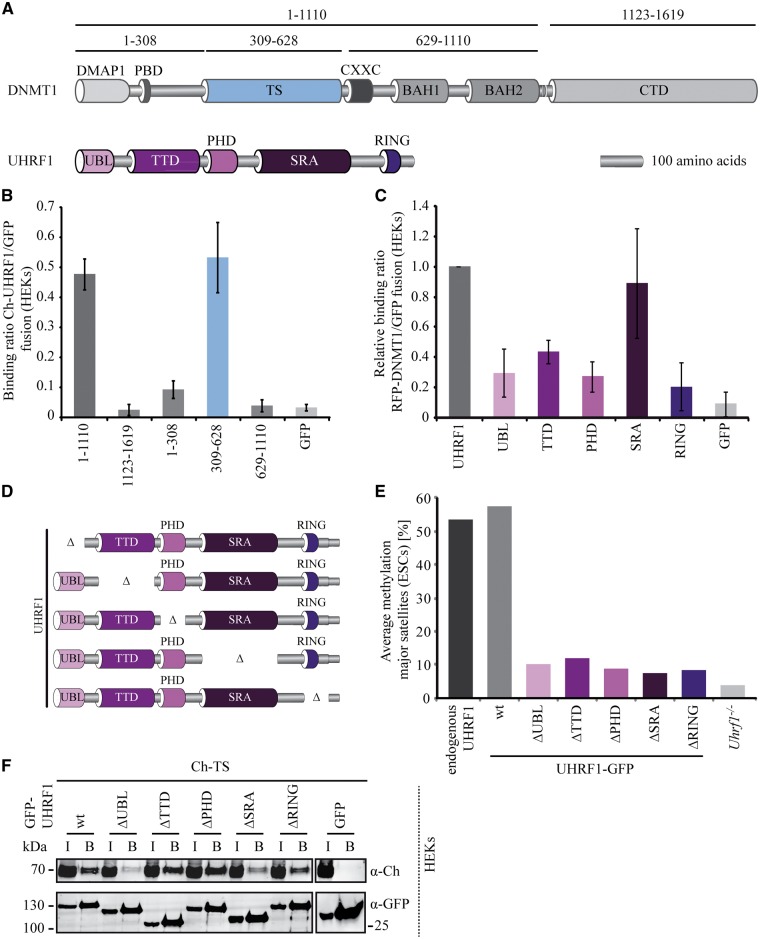
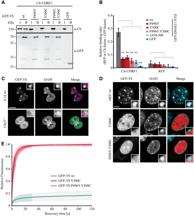
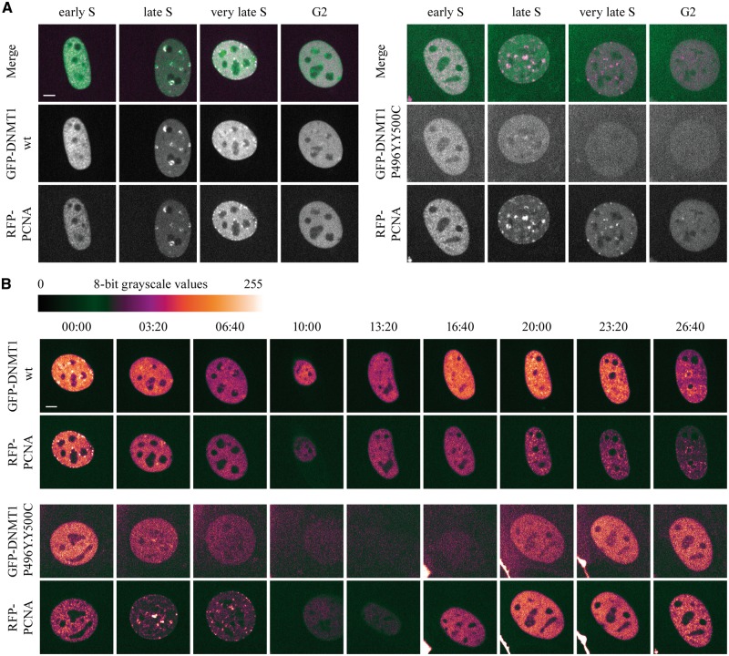
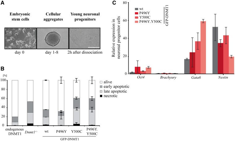
DNMT1 is recruited to substrate sites by PCNA and UHRF1 to maintain DNA methylation after replication. The cell cycle dependent recruitment of DNMT1 is mediated by the PCNA-binding domain (PBD) and the targeting sequence (TS) within the N-terminal regulatory domain. The TS domain was found to be mutated in patients suffering from hereditary sensory and autonomic neuropathies with dementia and hearing loss (HSANIE) and autosomal dominant cerebellar ataxia deafness and narcolepsy (ADCA-DN) and is associated with global hypomethylation and site specific hypermethylation. With functional complementation assays in mouse embryonic stem cells, we showed that DNMT1 mutations P496Y and Y500C identified in HSANIE patients not only impair DNMT1 heterochromatin association, but also UHRF1 interaction resulting in hypomethylation. Similar DNA methylation defects were observed when DNMT1 interacting domains in UHRF1, the UBL and the SRA domain, were deleted. With cell-based assays, we could show that HSANIE associated mutations perturb DNMT1 heterochromatin association and catalytic complex formation at methylation sites and decrease protein stability in late S and G2 phase. To investigate the neuronal phenotype of HSANIE mutations, we performed DNMT1 rescue assays and could show that cells expressing mutated DNMT1 were prone to apoptosis and failed to differentiate into neuronal lineage. Our results provide insights into the molecular basis of DNMT1 dysfunction in HSANIE patients and emphasize the importance of the TS domain in the regulation of DNA methylation in pluripotent and differentiating cells.


Figures

Figure 1
Figure 1
Domain structure of mouse DNMT1 and conservation of the TS domain among different species. (A) Domain structure of GFP-DNA methyltransferase 1 (DNMT1) with illustration of deletions and HSANIE associated point mutations in the targeting sequence (TS) domain. The large regulatory N-terminal domain (NTD) of DNMT1 is comprised of a DNA methyltransferase associated protein 1 (DMAP1)-binding domain, a proliferating cell nuclear antigen (PCNA)-binding domain (PBD), the TS domain harboring a ubiquitin interacting motif (UIM (21)), a zinc finger (CXXC) domain, two bromo-adjacent homology (BAH1 and BAH2) domains and a catalytically active C-terminal domain (CTD). (B) Primary sequence alignment of TS domains from different species. The secondary structure of the mouse TS domain is indicated (PDB: 3AV4 (23)). Highly conserved residues are shaded black. Deleted regions are indicated by blue rectangles and hereditary sensory and autonomic neuropathy type IE (HSANIE) associated point mutations by red arrows.
Figure 2
Figure 2
TS domain deletions and HSANIE associated mutations affect the DNA methylation activity of DNMT1 in vivo. (A and B) Site-specific DNA methylation analysis at major satellite repeats of mouse Dnmt1−/− ESCs expressing transiently GFP-DNMT1 wild-type (wt) and deletions (A) or HSANIE associated point mutant constructs (B). DNA methylation levels of untransfected Dnmt1−/− ESCs are displayed for comparison. Shown are mean values ± standard deviations (SDs) from two to six (A) or (B) three independent biological replicates (average from eight CpG sites, respectively). (B) Two-sample t-tests were performed that assume equal variances. Asterisks represent significant differences: *P < 0.05, **P < 0.001. (C) Quantitative evaluation of FRAP data showing mean curves. Error bars represent standard deviations. FRAP of S-phase mouse embryonic fibroblasts (n = 5) expressing GFP-DNMT1 and GFP-DNMT1 harboring HSANIE mutations without drug treatment (upper panel) and after treatment with 30 µM 5-aza-dC for 30 min (lower panel).
Figure 3
Figure 3
DNMT1 TS domain interaction with UHRF1 is necessary for DNA methylation. (A) Schematic outline of DNMT1 and the ubiquitin-like, containing PHD and RING finger domains 1 (UHRF1) expression constructs used for protein–protein interaction mapping studies. UHRF1 harbors an ubiquitin-like domain (UBL) followed by a tandem Tudor domain (TTD), a plant homeodomain (PHD), a SET and RING associated (SRA) domain and a really interesting new gene (RING) domain. (B) Mapping and relative quantification of the interaction GFP-DNMT1 with Ch-UHRF1 by fluorescence protein–protein interaction assay in vitro. Ratios of Ch-UHRF1 over GFP fusion proteins are shown as mean values ± standard error of the mean (SEM) of three to six biological replicates. (C) Mapping and relative quantification of the interaction GFP-UHRF1 with RFP-DNMT1 by fluorescence protein–protein interaction assay in vitro. Ratios of RFP-DNMT1 over GFP fusion proteins are shown as mean values ± SEM of three biological replicates normalized to the binding ratio of the GFP-UHRF1 full length protein. (D) Schematic outline of different UHRF1-GFP single domain deletion (Δ) constructs used for rescue experiments. (E) Local methylation analysis at major satellite repeats. CpG methylation levels in Uhrf1−/− ESCs stably expressing UHRF1-GFP wt or single domain deletions were analyzed. DNA methylation levels of untransfected Uhrf1−/− ESCs are shown for comparison. Values represent means from eight CpG sites. (F) Coimmunoprecipitation of UHRF1-GFP wt and single domain deletion mutants and Ch-TS. GFP and Ch fusion constructs were coexpressed in HEK293T cells and cell lysates were used for immunoprecipitation with the GFP-Trap. Bound fractions were detected by immunoblotting with anti-GFP and anti-Ch antibodies. GFP was used as negative control. I = Input, B = Bound.
Figure 4
Figure 4
HSANIE associated point mutations in the TS domain reduce the interaction of DNMT1 with UHRF1. (A) Coimmunoprecipitation of GFP-TS wt, P496Y, Y500C and P496Y.Y500C mutant constructs and Ch-UHRF1. GFP and Ch fusion constructs were coexpressed in HEK293T cells and cell lysates were used for immunoprecipitation with the GFP-Trap. Bound fractions were analyzed by immunoblotting with anti-GFP and anti-Ch antibodies. GFP was used as negative control. I = Input, B = Bound. (B) Fluorescence protein–protein interaction assay of the GFP-DNMT1 NTD with Ch-UHRF1. After one-step purification of the GFP-DNMT1 NTD wt and mutant constructs in a GFP-multiTrap plate, the binding of Ch-UHRF1 expressed in HEK293T cells was determined by fluorescence readout. GFP and RFP were used as negative control. Shown are mean relative binding ratios ± SEM of Ch-UHRF1 or RFP over GFP fusion proteins from four to six biological replicates. Two-sample t-tests were performed that assume equal variances. Significance compared to the relative binding ratio of GFP-DNMT1 NTD wt are indicated: *P < 0.05, **P < 0.001. (C) Confocal midsections of fixed mouse ESCs (wt, Uhrf1−/−) transiently expressing GFP-TS and DNA was counterstained with DAPI. In the merged image, DAPI is depicted in magenta. Scale bar 10 µm; enlargements: 3-times magnification, scale bar 2 µm. (D) Confocal midsections of fixed MEF cells transfected with GFP-TS wt or GFP-TS Y500C and P496Y.Y500C constructs. In the merged image, GFP-TS is depicted in red and DAPI in magenta. Scale bar 5 µm; enlargements: 3-times magnification, scale bar 1 µm. (E) Protein mobility of GFP-TS wt and HSANIE associated GFP-TS Y500C and P497Y.Y500C mutants in living MEF cells (n = 13) determined by half nucleus fluorescent recovery after photobleaching (FRAP) analysis. Curves represent mean ± SEM.
Figure 5
Figure 5
The destabilization of the DNMT1 HSANIE associated mutations is cell cycle dependent. (A and B) Spinning disk confocal midsections of MEF cells transiently coexpressing GFP-DNMT1 wt or P496Y.Y500C mutant and RFP-PCNA as a cell cycle marker. (A) Selected frames from live cell series are shown of GFP-DNMT1 wt (left panel) and GFP-DNMT1 P496Y.Y500C (right panel). Cells were tracked starting from early S until G2 phase. In the merged image, RFP-PCNA is depicted in magenta. (B) Live cell series of MEF cells shown in (A) transiently coexpressing GFP-DNMT1 wt (upper panel) or P496Y.Y500C (lower panel). Starting from very late S phase (wt) or mid S phase (P496Y.Y500C) images were taken every 200 min. White represents the highest and black the lowest intensity. Scale bar 5 µm.
Figure 6
Figure 6
HSANIE associated point mutations in the TS domain show different survival rate and differentiation potential during neuronal progenitor differentiation. (A) Mouse embryonic stem cells are differentiated into young neuronal progenitor cells (NPCs). Scale bar: 200 µm. (B) Flow cytometric analysis of alive, early apoptotic, late apoptotic and necrotic cells. Two hours after dissociation of cellular aggregates, young neuronal progenitors were analyzed using Annexin V and propidium iodide staining. Bar graphs represent mean values ± standard deviation (SD) from two to three biological replicates. (C) RNA expression profiles of NPCs in biological duplicates (wt) or triplicates (P496Y, Y500C, P496Y.Y500C) of pluripotency factor Oct4, lineage specific markers Brachyury, Gata6 and Nestin for mesodermal, endodermal and ectodermal lineage, respectively. All ddCt values are normalized to wt Oct4 expression.

Similar articles

Cited by

References

    1. Bird A. (2002) DNA methylation patterns and epigenetic memory. Genes Dev., 16, 6–21. - PubMed
    1. Goll M.G., Bestor T.H. (2005) Eukaryotic cytosine methyltransferases. Annu. Rev. Biochem., 74, 481–514. - PubMed
    1. Cheng X., Blumenthal R.M. (2008) Mammalian DNA methyltransferases: a structural perspective. Structure, 16, 341–350. - PMC - PubMed
    1. Law J.A., Jacobsen S.E. (2010) Establishing, maintaining and modifying DNA methylation patterns in plants and animals. Nat. Rev. Genet., 11, 204–220. - PMC - PubMed
    1. Smith Z.D., Meissner A. (2013) DNA methylation: roles in mammalian development. Nat. Rev. Genet., 14, 204–220. - PubMed

MeSH terms

Substances