. 2017 Jun 2;45(10):5707-5719.
doi: 10.1093/nar/gkx142.

Structural and mechanistic insights into regulation of HBO1 histone acetyltransferase activity by BRPF2

Affiliations

Structural and mechanistic insights into regulation of HBO1 histone acetyltransferase activity by BRPF2

Ye Tao et al. Nucleic Acids Res. .

Abstract

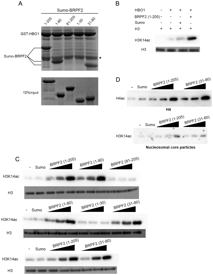
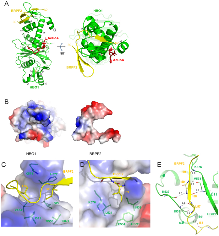
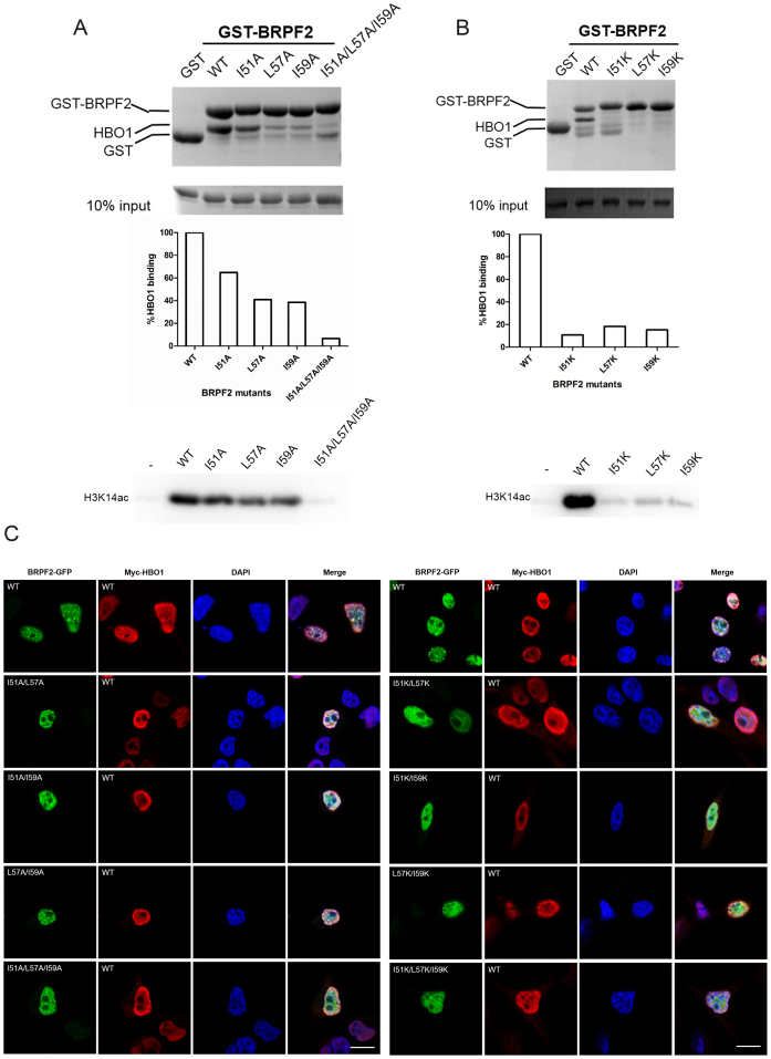
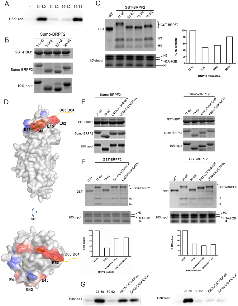
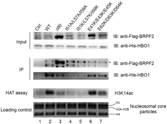
HBO1, a member of the MYST family of histone acetyltransferases (HATs), is required for global acetylation of histone H3K14 and embryonic development. It functions as a catalytic subunit in multisubunit complexes comprising a BRPF1/2/3 or JADE1/2/3 scaffold protein, and two accessory proteins. BRPF2 has been shown to be important for the HAT activity of HBO1 toward H3K14. Here we demonstrated that BRPF2 can regulate the HAT activity of HBO1 toward free H3 and H4, and nucleosomal H3. Particularly, a short N-terminal region of BRPF2 is sufficient for binding to HBO1 and can potentiate its activity toward H3K14. The crystal structure of the HBO1 MYST domain in complex with this segment of BRPF2 together with the biochemical and cell biological data revealed the key residues responsible for the HBO1-BRPF2 interaction. Our structural and functional data together indicate that the N-terminal region of BRPF2 plays an important role in the binding of HBO1 and a minor role in the binding of nucleosomes, which provide new mechanistic insights into the regulation of the HAT activity of HBO1 by BRPF2.


Figures

Figure 1.
Figure 1.
BRPF2 can potentiate the HAT activity of HBO1 towards H3K14 in vitro. (A) In vitro GST pull-down assay of the GST-tagged HBO1 MYST domain with different Sumo-tagged BRPF2 fragments. Equal loading of BRPF2 is shown. The band corresponding to the GST tag band is indicated by an asterisk. (B) Potentiation of the HAT activity of the HBO1 MYST domain by the N-terminal region of BRPF2 (1–205). In vitro HAT activity assay was performed on free full-length H3. The acetylation levels of H3 were analyzed with an antibody highly specific to acetylated H3K14 using half of the reaction mixture. Equal loading of H3 is shown with Coomassie blue staining of the other half of the reaction mixture. (C) In vitro HAT activity assay of the HBO1 MYST domain towards H3K14 in the presence of different Sumo–BRPF2 fragments. The molar ratio of BRPF2 and HBO1 was set at 0.5:1, 1:1 and 2:1, respectively. Equal loading of H3 is shown as in (B). (D) In vitro HAT activity assay of the HBO1 MYST domain toward free full-length H4 and nucleosomal H3 in the presence of Sumo–BRPF2 (1–205) and Sumo–BRPF2 (31–80).
Figure 2.
Figure 2.
Structure of the HBO1–BRPF2 complex. (A) A ribbon representation of the overall structure of the HBO1–BRPF2 complex. The HBO1 MYST domain and the BRPF2 (residues 39–62) fragment are colored in green and yellow, respectively. The bound AcCoA and Zn2+ are shown with a red stick model and a gray sphere, respectively. (B) Electrostatic surface representations of HBO1 and BRPF2 showing good complementarity in both geometrical and electrostatic properties. (C and D) Hydrophobic interactions between HBO1 and BRPF2. HBO1 is shown with an electrostatic surface and BRPF2 is shown with a ribbon diagram. The residues involved in the hydrophobic interactions are shown with side chains. (E) Hydrophilic interactions between HBO1 and BRPF2. Both HBO1 and BRPF2 are shown with ribbon diagrams. The hydrophilic interactions are indicated with dotted lines and distances.
Figure 3.
Figure 3.
Validation of the functional roles of key residues of BRPF2. (A and B) In vitro functional analyzes of BRPF2 (31–80) mutants. BRPF2 (31–80) mutants with mutations of Ile51, Leu57 and/or Ile59 to (A) alanine or (B) lysine were analyzed. In vitro GST pull-down assays of wild-type (WT) and mutant GST–BRPF2 (31–80) with the HBO1 MYST domain were performed. Equal loading of HBO1 is shown. The amount of HBO1 that was pulled down was semi-quantitated. In vitro HAT activity assays of HBO1 toward H3K14 in the presence of WT or mutant Sumo–BRPF2 (31–80) were performed, and the acetylated H3K14 was detected with a specific antibody. (C) Localization of HBO1 and BRPF2 in transfected HEK 293T cells. Cells were co-transfected with full-length Myc-HBO1 and full-length WT or mutant BRPF2–GFP harboring mutations to alanine (left panel) or lysine (right panel). Immunofluorescence with anti-Myc antibody was performed to detect HBO1. Localization of HBO1 and BRPF2 was examined with confocal fluorescence microscopy. Scale bar represents 10 μm.
Figure 4.
Figure 4.
Examination of potential binding of the N-terminal region of BRPF2 with histones in vitro. (A) In vitro HAT activity assays of HBO1 toward H3K14 in the presence of different Sumo–BRPF2 fragments. (B) In vitro GST pull-down assays of GST–HBO1 with different Sumo–BRPF2 fragments. Equal loading of Sumo–BRPF2 is shown. (C) In vitro GST pull-down assays of different GST–BRPF2 fragments with free H3 and H4 from histone mixture. The amount of histone H3 that was pulled down was semi-quantitated. (D) Two negatively charged surface patches in the N-terminal region of BRPF2. HBO1 is shown with gray surface. BRPF2 is shown with electrostatic surface and the conserved acidic residues are shown with side chains. Asp63 and Asp64 are invisible in the HBO1–BRPF2 structure and thus are indicated with a dash line. (E) In vitro GST pull-down assays of GST–HBO1 with WT or mutant Sumo–BRPF2 (31–80). Equal loading of Sumo–BRPF2 is shown. (F) In vitro GST pull-down assays of WT or mutant GST–BRPF2 (31–80) with free H3 and H4 from histone mixture. The amount of histone H3 that was pulled down was semi-quantitated. (G) In vitro HAT activity assays of HBO1 toward H3K14 in the presence of WT or mutant Sumo–BRPF2 (31–80).
Figure 5.
Figure 5.
Examination of the functional role of the N-terminal region of BRPF2 in vivo. HEK 293T cells were co-transfected with full-length His-HBO1 and WT or mutant Flag-BRPF2, and cells transfected with His-HBO1 alone served as the negative control. Immunoprecipitation (IP) experiments were performed with nuclear extracts of the transfected cells using an anti-Flag antibody. The IPed BRPF2–HBO1 complexes were then subjected to HAT activity assays using NCPs as the substrate. The nuclear extracts, the IPed complexes and the reaction mixture of the HAT activity assays were analyzed with sodium dodecyl sulphate-polyacrylamide gel electrophoresis (SDS-PAGE) and western blotting using antibodies specific to the Flag tag, His tag, and acetylated H3K14. The band corresponding to the full-length Flag-BRPF2 protein was denoted by an asterisk and the band below might represent the degraded Flag-BRPF2 protein. Equal loading of NCPs in the HAT activity assays were shown with SDS-PAGE and Coomassi blue staining.

Similar articles

Cited by

References

    1. Suganuma T., Workman J.L.. Signals and combinatorial functions of histone modifications. Annu. Rev. Biochem. 2011; 80:473–499. - PubMed
    1. Bannister A.J., Kouzarides T.. Regulation of chromatin by histone modifications. Cell Res. 2011; 21:381–395. - PMC - PubMed
    1. Marmorstein R., Zhou M.M.. Writers and readers of histone acetylation: structure, mechanism, and inhibition. Cold Spring Harb. Perspect. Biol. 2014; 6:a018762. - PMC - PubMed
    1. Tessarz P., Kouzarides T.. Histone core modifications regulating nucleosome structure and dynamics. Nat. Rev. Mol. Cell Biol. 2014; 15:703–708. - PubMed
    1. Sapountzi V., Cote J.. MYST-family histone acetyltransferases: beyond chromatin. Cell Mol. Life Sci. 2011; 68:1147–1156. - PMC - PubMed

MeSH terms

LinkOut - more resources