. 2017 May 15;26(10):1863-1876.
doi: 10.1093/hmg/ddx090.

Amelogenesis imperfecta caused by N-terminal enamelin point mutations in mice and men is driven by endoplasmic reticulum stress

Affiliations

Amelogenesis imperfecta caused by N-terminal enamelin point mutations in mice and men is driven by endoplasmic reticulum stress

Steven J Brookes et al. Hum Mol Genet. .

Abstract

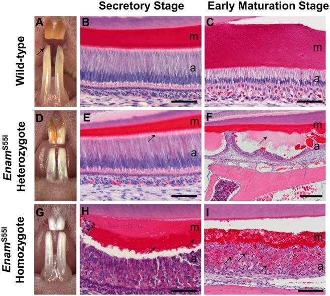
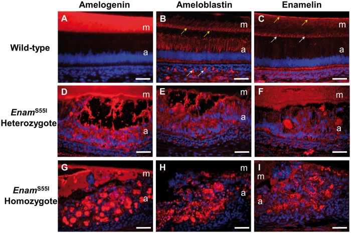
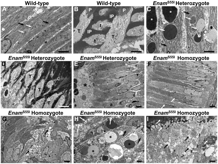
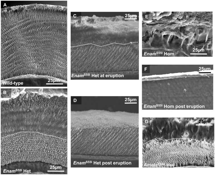
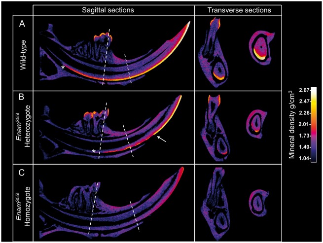
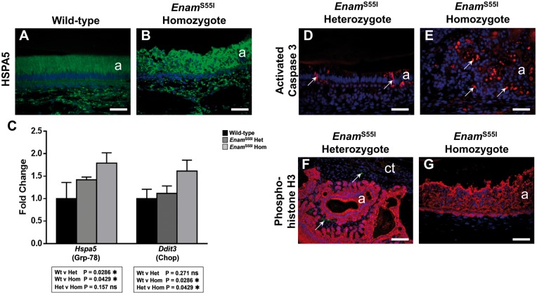
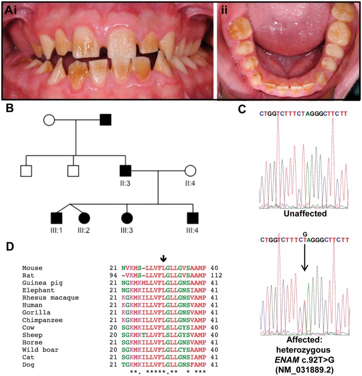
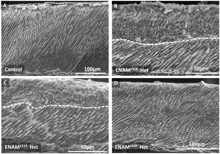
'Amelogenesis imperfecta' (AI) describes a group of inherited diseases of dental enamel that have major clinical impact. Here, we identify the aetiology driving AI in mice carrying a p.S55I mutation in enamelin; one of the most commonly mutated proteins underlying AI in humans. Our data indicate that the mutation inhibits the ameloblast secretory pathway leading to ER stress and an activated unfolded protein response (UPR). Initially, with the support of the UPR acting in pro-survival mode, Enamp.S55I heterozygous mice secreted structurally normal enamel. However, enamel secreted thereafter was structurally abnormal; presumably due to the UPR modulating ameloblast behaviour and function in an attempt to relieve ER stress. Homozygous mutant mice failed to produce enamel. We also identified a novel heterozygous ENAMp.L31R mutation causing AI in humans. We hypothesize that ER stress is the aetiological factor in this case of human AI as it shared the characteristic phenotype described above for the Enamp.S55I mouse. We previously demonstrated that AI in mice carrying the Amelxp.Y64H mutation is a proteinopathy. The current data indicate that AI in Enamp.S55I mice is also a proteinopathy, and based on comparative phenotypic analysis, we suggest that human AI resulting from the ENAMp.L31R mutation is another proteinopathic disease. Identifying a common aetiology for AI resulting from mutations in two different genes opens the way for developing pharmaceutical interventions designed to relieve ER stress or modulate the UPR during enamel development to ameliorate the clinical phenotype.


Figures

Figure 1
Figure 1
Gross and histological appearance of the incisor teeth of wild-type, EnamS55I heterozygous and EnamS55I homozygous mice. (A) The teeth of wild-type mice have a smooth, orange-coloured, translucent appearance with a sharp incisal edge (arrow). (B) Histologically, during the secretory stage of amelogenesis, the enamel organ is composed of tall, columnar ameloblasts which secrete an eosinophilic matrix. (C) During the early maturation stage of amelogenesis the ameloblasts have shortened considerably and the matrix has begun to degrade (less intense eosin staining). (D) The teeth of EnamS55I heterozygous mice are largely white with patches of normal, orange-coloured enamel on their surface. (E) The secretory stage ameloblasts (a) are tall and columnar as in wild-type mice but their cytoplasm is more deeply eosin-stained. Contact of the ameloblasts with the matrix is more diffuse (arrow) than in their wild-type counterparts and the secreted matrix is considerably thinner. (F) By the early maturation stage, the ameloblasts have frequently lost contact with the matrix forming large blister-like structures (arrow) which contain pale-staining material. (G) The teeth of EnamS55I homozygous mice are completely white and devoid of enamel. (H) The ameloblasts have lost their characteristic columnar structure and form eosinophilic multicellular masses. The matrix produced by these cells is disorganized and contains cellular debris (arrows). (I) By the early maturation stage, the ameloblasts continue to form multicellular aggregates enclosing eosinophilic material (arrows) while the matrix appears thin and disorganized. a: ameloblasts; m; matrix. Scale bars: (B, C, E, H) 50 μm; (I), 100 μm; (F), 500 μm.
Figure 2
Figure 2
Enamel matrix protein distribution in EnamS55I mutant mice. (A–C) Wild-type mice. (A) Strong amelogenin immunoreactivity in the extracellular matrix of wild-type mice. (B) Ameloblastin immunoreactivity can be seen within the ameloblasts and at the secretory front (yellow arrows) of the matrix and extending as parallel lines into it. There is non-specific immunolabelling of the vasculature below the ameloblasts (white arrows). (C) Enamelin shows strong immunoreactivity at the dentino-enamel junction (yellow arrows) and at the secretory front (white arrows) with less intense immunostaining throughout the remainder of the matrix. (D–F) EnamS55I heterozygous mice. (D) In EnamS55I heterozygous mice there is strong amelogenin immunoreactivity in the matrix and within the ameloblasts. (E) Little immunoreactivity is observed in the matrix of heterozygous mice but there is strong intracellular immunolabelling for ameloblastin in the ameloblasts. (F) Strong enamelin immunoreactivity is seen throughout the matrix and within the ameloblasts. (G–I) EnamS55I homozygous mice. (G) There is intense amelogenin immunoreactivity both in the matrix and within the ameloblasts. (H) Strong ameloblastin immunolabelling is confined to the ameloblasts. (I) There is strong enamelin immunoreactivity within the ameloblasts and weaker immunostaining of the matrix. a: ameloblasts; m; matrix. Scale bars: 50 μm.
Figure 3
Figure 3
Ultrastructure of EnamS55I ameloblasts. (A, B) Wild-type secretory stage ameloblasts (A) display a tall columnar morphology with abundant, peripherally organized rough endoplasmic reticulum (black arrows) surrounding a centrally placed Golgi apparatus and associated small vesicles (white arrows). (B) At the ameloblast tip is a specialized secretory structure, the Tomes’ process (T) that secretes enamel matrix in such a manner that the matrix that is deposited from the tip gives rise to enamel prisms (P) while that derived from its flanks produces inter-prismatic (IP) enamel. Adjacent Tomes’ processes, sectioned more transversely, can also be seen and these secrete their enamel matrix in a different orientation, (t), to give rise to the interlocking arrangement of enamel prisms. (C–E)EnamS55I heterozygous mice the secretory ameloblasts also have (C) abundant rough endoplasmic reticulum (black arrows) but also contain larger intracellular vesicles of varying electron translucency (white arrows) when compared to wild-type. Extracellular accumulation of matrix-like material is also seen just under the secretory front of the ameloblasts (white asterisks). (D) The Tomes’ processes of these ameloblasts are abnormal (T) appearing disorganized, crowded and slim. (E) As the maturation stage of amelogenesis is approached, the ameloblasts (a) of EnamS55I heterozygous mice retract their Tomes’ processes and lose contact with the enamel matrix and form cyst-like structures (black asterisk) containing matrix-like material. (F–I) The secretory stage ameloblasts of EnamS55I homozygous mice are initially columnar (F) with accumulations of matrix-like material between them (white arrows). (G) Later in the secretory stage of amelogenesis, EnamS55I homozygous ameloblasts lose their columnar morphology and become ovoid or spherical. Many of the cells contain large vacuoles (black arrows). (H) At higher magnification the ameloblasts of EnamS55I homozygous mice contain prominent rough endoplasmic reticulum (black arrows). Large vesicles containing material of strong to weak electron translucency (black asterisks) as well as vacuolar structures (white arrows) are often seen in these ameloblasts. (I) At later stages of amelogenesis, homozygous ameloblasts show extensive vacuolation (black arrows) and accumulations of matrix like material appear around and between them (white arrows). Scale bars: (A, B, C, D), 2 μm; (E, F, G), (I) 5 μm; (H) 1 μm.
Figure 4
Figure 4
SEM comparing the structure of wild-type, heterozygous and homozygous EnamS55I and female heterozygous AmelxY64H mandibular incisor enamel. (A) Transverse sections through a wild-type incisor at eruption show the well organized, decussating pattern of enamel prisms characteristic of rodent incisor enamel. Only the very outermost layer of enamel is aprismatic. (B) In contrast, heterozygous EnamS55I enamel exhibits a structurally normal decussating inner layer of enamel topped with a subsequently secreted layer that is structurally abnormal. A dotted white line marks the boundary between the two layers. (C) Sagittal sections through heterozygous EnamS55I enamel at the point of eruption show that a structurally abnormal outer enamel layer is present. (D) Later, in post-eruptive enamel nearer the incisal biting edge, the abnormal layer has been lost and only the inner structurally normal enamel layer survives. (E) Homozygous EnamS55I mice fail to secrete any tissue with structural resemblance to normal enamel (unerupted transverse section). (F) Post-eruptive homozygous EnampS55I incisors exhibit no enamel layer; just a thin layer of material of unknown composition. (G) The phenotype of heterozygous AmelxpY64H mouse enamel (39) is remarkably similar to that of heterozygous EnampS55I mice; both exhibiting a structurally normal inner layer and an abnormal outer layer.
Figure 5
Figure 5
Calibrated micro X-ray computed tomography comparing mineral density in wild-type, heterozygous and homozygous EnamS55I enamel. (A) Sagittal CT sections show that wild-type incisor enamel becomes X-ray opaque due to increased mineral density (secondary mineralization) that occurs at the beginning of the maturation stage. Mineral density increases gradually throughout maturation and following eruption. Transverse CT sections, taken at points indicated by the dotted lines, show the enamel layer is complete across the width of the labial aspect of the incisor. (B) In contrast, onset of secondary mineralization is delayed in heterozygous EnamS55I enamel (asterisk) and only approaches the density seen in wild-type mice following eruption. Transverse sections show that secondary mineralization is compromised across the whole width of the enamel. (C) Homozygous EnamS55I do not exhibit any X-ray opaque material corresponding to incisor enamel. Only wild-type and heterozygous EnamS55I mice exhibit molar enamel (A and B). The molar teeth of all mice are fully erupted in these mice and no developing enamel is present.
Figure 6
Figure 6
Analysis of cell death, proliferation and ER stress in EnamS55I mutant mice. (A, B) Immunolabelling for the ER chaperone HSPA5 (Grp-78) showed moderate immunoreactivity in wild-type secretory stage ameloblasts (A) and also in those of EnamS55I heterozygous mice (data not shown). The ameloblasts of EnamS55I homozygous mice (B) showed a substantial increase in HSPA5 immunofluorescence. (C) Quantitative reverse transcriptase PCR analysis of ER stress gene expression in the secretory stage enamel organ. Hspa5 (Grp-78) gene expression was statistically significantly elevated in EnamS55I heterozygous and homozygous mice compared to wild-type. Ddit3 (Chop) gene expression, was statistically significantly elevated in EnamS55I homozygous mice but not in EnamS55I heterozygous secretory stage enamel organ compared to wild-type. The data was analysed using the one-sided Mann-Whitney test. (D, E) Immunolabelling for activated caspase 3 in EnamS55I heterozygous ameloblasts (D) shows occasional apoptotic ameloblasts at the transition stage of amelogenesis (arrows) as is typically seen in wild-type animals (data not shown). In EnamS55I homozygous mice, many of the aberrant ameloblasts in the multicellular masses were seen to be undergoing apoptosis (arrows) (E). (F, G) Dual immunolabelling for phosphohistone H3 (green fluorescence), a marker of cell proliferation and keratin 14 (red fluorescence), to discriminate the enamel organ from the surrounding connective tissue, showed that in all mutant mice studied none of the ameloblast population showed evidence of proliferation. (F) Where proliferation was observed, this was confined to the surrounding connective tissue fibroblast population (arrows). Scale bars: (A, B, D, E, G), 50 μm; (F), 100 μm.
Figure 7
Figure 7
(Ai and ii) The permanent dentition of III:4 is characterized by a generalized rough hypoplastic AI with superficial exogenous staining. (B) Pedigree of family 1. Individuals are not necessarily shown in age order. Affected individuals are indicated by shading. DNA was available from labelled individuals. Whole exome sequencing was carried out using DNA from III:2. Teeth were obtained from III:1 and III:4. (C) Sanger sequencing of ENAM exon 3 confirmed the heterozygous c.92T > G variant (NM_031889.2), originally identified in III:2 by WES, segregated with the disease phenotype in all available family members. (D) Clustal Omega multiple sequence alignment of homologous ENAM protein sequences. The arrow indicates the residue altered by the c.92T > G variant, p.L31R (NM_031889.2; NP_114095.2). The affected residue and the ten residues that flank it to either side are included in the alignment. ENAM sequences used: Mouse, Mus musculus NP_059496.1; Rat Rattus norvegicus NP_001099471.1; Guinea pig, Cavia porcellus XP_003467585.2; Elephant, Loxodonta africana XP_003414215.1; Rhesus macaque, Macaca mulatta XP_014994062.1; Human, Homo sapiens NP_114095.2; Gorilla, Gorilla gorilla XP_004038829.1; Chimpanzee, Pan troglodytes XP_526591.1; Cow, Bos taurus XP_605463.4; Sheep Ovis aries XP_004009936.1; Horse Equus caballus XP_001487944.1; Wild boar, Sus scrofa NP_999406.1; Cat, Felis Catus XP_003985351.1; Dog Canis lupus familiaris XP_539305.3.
Figure 8
Figure 8
SEM comparison between human control primary incisor enamel and primary incisor enamel suffering the effects of the heterozygous ENAMpL31R mutation. (A) Control enamel exhibits the typical prismatic structure with organized prisms spanning the entire enamel thickness. (B, C) In contrast, affected enamel recapitulates the phenotype seen in heterozygous EnampS55I and AmelxpY64H mice in that it exhibits an inner layer of enamel exhibiting ordered prismatic structure overlaid with an outer enamel layer that is structurally abnormal (the two layers being demarcated by the white dotted line). (D) In some areas, affected enamel appears structurally normal and does not exhibit a structurally abnormal outer layer. However, it is not clear whether these apparently unaffected areas simply represent areas where the structurally abnormal outer layer has been lost due to wear and tear experienced in the mouth.

Similar articles

Cited by

References

    1. Nanci A. (2007) Ten Cate's Oral Histology: Development, Structure And Function. Mosby, St. Louis, MO.
    1. Bartlett J.D., Ganss B., Goldberg M., Moradian-Oldak J., Paine M.L., Snead M.L., Wen X., White S.N., Zhou Y.L. (2006) 3. Protein-protein interactions of the developing enamel matrix. Curr. Top. Dev. Biol., 74, 57–115. - PubMed
    1. Smith C.E. (1998) Cellular and chemical events during enamel maturation. Crit. Rev. Oral Biol. Med., 9, 128–161. - PubMed
    1. Backman B., Holm A.K. (1986) Amelogenesis imperfecta: prevalence and incidence in a northern Swedish county. Commun. Dent. Oral Epidemiol., 14, 43–47. - PubMed
    1. Coffield K.D., Phillips C., Brady M., Roberts M.W., Strauss R.P., Wright J.T. (2005) The psychosocial impact of developmental dental defects in people with hereditary amelogenesis imperfecta. J. Am. Dent. Assoc., 136, 620–630. - PubMed

Publication types