. 2017 Apr 20;45(7):3953-3966.
doi: 10.1093/nar/gkx166.

Handcuffing reversal is facilitated by proteases and replication initiator monomers

Affiliations

Handcuffing reversal is facilitated by proteases and replication initiator monomers

Katarzyna Bury et al. Nucleic Acids Res. .

Abstract

Specific nucleoprotein complexes are formed strictly to prevent over-initiation of DNA replication. An example of those is the so-called handcuff complex, in which two plasmid molecules are coupled together with plasmid-encoded replication initiation protein (Rep). In this work, we elucidate the mechanism of the handcuff complex disruption. In vitro tests, including dissociation progress analysis, demonstrate that the dimeric variants of plasmid RK2 replication initiation protein TrfA are involved in assembling the plasmid handcuff complex which, as we found, reveals high stability. Particular proteases, namely Lon and ClpAP, disrupt the handcuff by degrading TrfA, thus affecting plasmid stability. Moreover, our data demonstrate that TrfA monomers are able to dissociate handcuffed plasmid molecules. Those monomers displace TrfA molecules, which are involved in handcuff formation, and through interaction with the uncoupled plasmid replication origins they re-initiate DNA synthesis. We discuss the relevance of both Rep monomers and host proteases for plasmid maintenance under vegetative and stress conditions.


Figures

Figure 1.
Figure 1.
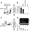
RK2 plasmid handcuff complex is formed by wild-type TrfA. Schematic representation of the experimental procedure is presented in panel (A) NA assay, and (C) ligation assay. The formation of handcuff complexes was analyzed using the following proteins: wild-type TrfA, monomeric form TrfA G254D/S267L and dimeric form TrfA S257F, as described under Materials and methods. The results of performed experiments are presented in panel (B) NA assay, and (D) ligase assay. On agarose gel free DNA and DNA multimers are marked F and M respectively. A quantified densitometry with using ChemiDoc MP System and ImageLab™ Software (BioRad) were applied for DNA multimers amount estimation. The error bars are derived from three different experiments.
Figure 2.
Figure 2.
RK2 iteron plasmid forms stable handcuff structures. The scheme of the experimental procedure is presented in panel (A). Stability of the RK2 handcuff structures was tested in the NA assay (B), on pre-assembled handcuff structures, as described under Materials and methods. Stability test was performed in continuous buffer rinsing. The fluorescence signals of particular samples, were analyzed after 0, 5, 15, 30, 45 and 60 min of buffer rinsing.
Figure 3.
Figure 3.
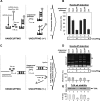
Uncuffing of the RK2 handcuff structures is mediated by Lon and ClpAP proteases. The scheme of the experimental procedure is presented in panel (A) NA assay, and (C) ligation assay. Uncuffing of the RK2 handcuff structures was tested on pre-assembled handcuff structures as described in Materials and methods. After handcuffing reaction the influence of several chaperones (ClpX and ClpA) and proteases (ClpXP, ClpAP and Lon) was analyzed by the NA (B) and ligase (D) assays. On agarose gel free DNA and DNA multimers are marked F and M, respectively. Positive control (lane 1, B and D) reactions with wild-type TrfA protein without the use of chaperones or proteases. Stability of TrfA protein, during analysis of handcuff structure disruption by chaperones and proteases in ligase assays, was tested by SDS-PAGE electrophoresis (E). A quantified densitometry with using ChemiDoc MP System and ImageLab™ Software (BioRad) were applied for TrfA protein and DNA multimers amount estimation. The error bars are derived from three different experiments.
Figure 4.
Figure 4.
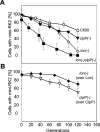
The mutation in Lon protease in E. coli strains affects the miniRK2 plasmid stability. Stability assays were performed as described under Materials and methods. The graphs show the dependence of the number of bacterial cells carrying plasmid pTJS42 on the number of generations after abolishment of the antibiotic selection pressure. Panel (A) shows the stability of mini-RK2 plasmid in E. coli C600 and its protease-deficient derivatives. Panel (B) shows the results of mini-RK2 plasmid stability experiments in Lon and ClpP supplemented strains. Cultures were grown for about 120 generations. The given results are means from three independent repeats of each experiment.
Figure 5.
Figure 5.
Monomeric form of TrfA protein uncuffs the RK2 handcuff structures. Schemes of the experimental procedure are presented in panel (A) NA assay, and (C) ligation assay. Uncuffing of the RK2 handcuff structures was tested on pre-assembled handcuff structures as described under Materials and methods. After handcuffing reaction the influence of monomeric TrfA form was analyzed in NA (B) and ligase (D) assays. On agarose gel free DNA and DNA multimers are marked F and M, respectively. A quantified densitometry with using ChemiDoc MP System and ImageLab™ Software (BioRad) were applied for DNA multimers amount estimation. The error bars are derived from three different experiments.
Figure 6.
Figure 6.
Monomeric form of the TrfA protein replaces the wild-type form from the plasmid DNA iterons. Schemes of the experimental procedure are presented in panel (A) handcuff formation with the fluorescently-labeled wild-type TrfA AlexaFluor 488, and (C) handcuff formation with the wild-type TrfA. After handcuffing reaction the influence of monomeric TrfA form was analyzed: (B) handcuff structures formed with TrfA AlexaFluor 488, were titrated with the monomeric form of TrfA protein (0.3 and 3 μM) and (D) handcuff structures formed with the wild-type TrfA protein were titrated with fluorescently-labeled monomeric form TrfA G254D/S267L AlexaFluor 488 (0.3 and 3 μM).
Figure 7.
Figure 7.
The TrfA dimers are stably associated within the handcuff complex. Schemes of the experimental procedure are presented in panel (A) handcuff formation with fluorescently-labeled wild-type TrfA AlexaFluor 488, and (C) handcuff formation with wild-type TrfA. After handcuffing reaction the dynamics of such structure was analyzed: (B) handcuff structures formed with TrfA AlexaFluor 488, were titrated with wild-type TrfA protein (0.3 and 3 μM) and (D) handcuff structures formed with the wild-type TrfA protein were titrated with fluorescently-labeled wild-type TrfA AlexaFluor 488 (0.3 and 3 μM).
Figure 8.
Figure 8.
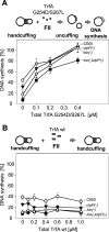
Monomeric form of TrfA protein re-initiates the RK2 plasmid replication. The influence of the monomeric (A) and wild-type form of TrfA protein (B), on handcuff structures, was tested in in vitro replication assay (FII). Reactions were performer using crude extracts obtained from E. coli C600 () or its protease-defective derivatives: clpP(−) (▾), lon(−) (⋄) and lon(−)clpP(−) (▪), as described under Materials and methods. All crude extracts were active for RK2 replication. Reactions contained 250 ng of supercoiled DNA template and 500 ng of wild-type TrfA protein. The starting points are the reactions in which, due to handcuffing, the maximum inhibition of DNA synthesis was observed. Formation of handcuff complexes was obtained through the addition of pBK20 iteron-containing plasmid in 2:1 molar ratio, in relation to pTJS42 RK2 mini-replicon. The influence of particular forms of TrfA protein was tested by titration of individual reactions by increasing concentration of monomeric TrfA (0.1, 0.2 and 0.4 μM) (A) or wild-type TrfA form (0.2, 0.4, 0.6 and 1.0 μM) (B). The error bars are derived from three repetitions of each reaction.

Similar articles

Cited by

References

    1. Ingmer H., Fong E.L., Cohen S.N.. Monomer-dimer equilibrium of the pSC101 RepA protein. J. Mol. Biol. 1995; 250:309–314. - PubMed
    1. Kolatka K., Kubik S., Rajewska M., Konieczny I.. Replication and partitioning of the broad-host-range plasmid RK2. Plasmid. 2010; 64:119–134. - PubMed
    1. Chattoraj D.K. Control of plasmid DNA replication by iterons: no longer paradoxical. Mol. Microbiol. 2000; 37:467–476. - PubMed
    1. Wickner S., Hoskins J., McKenney K.. Monomerization of RepA dimers by heat shock proteins activates binding to DNA replication origin. Proc. Natl. Acad. Sci. U.S.A. 1991; 88:7903–7907. - PMC - PubMed
    1. Ishiai M., Wada C., Kawasaki Y., Yura T.. Replication initiator protein RepE of mini-F plasmid: functional differentiation between monomers (initiator) and dimers (autogenous repressor). Proc. Natl. Acad. Sci. U.S.A. 1994; 91:3839–3843. - PMC - PubMed

MeSH terms

LinkOut - more resources