Case Reports
. 2017 May 1;26(9):1599-1611.
doi: 10.1093/hmg/ddx060.

A novel CISD2 mutation associated with a classical Wolfram syndrome phenotype alters Ca2+ homeostasis and ER-mitochondria interactions

Affiliations
Case Reports

A novel CISD2 mutation associated with a classical Wolfram syndrome phenotype alters Ca2+ homeostasis and ER-mitochondria interactions

Cécile Rouzier et al. Hum Mol Genet. .

Erratum in

Abstract

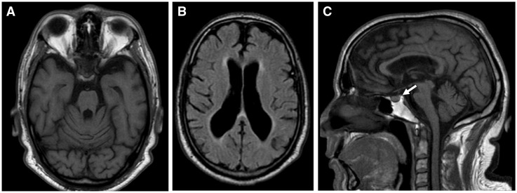
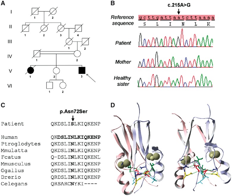
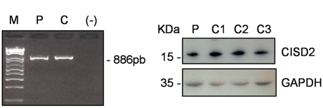
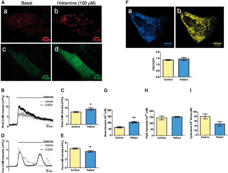
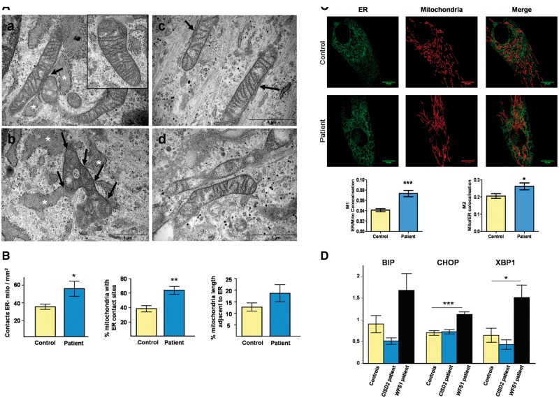
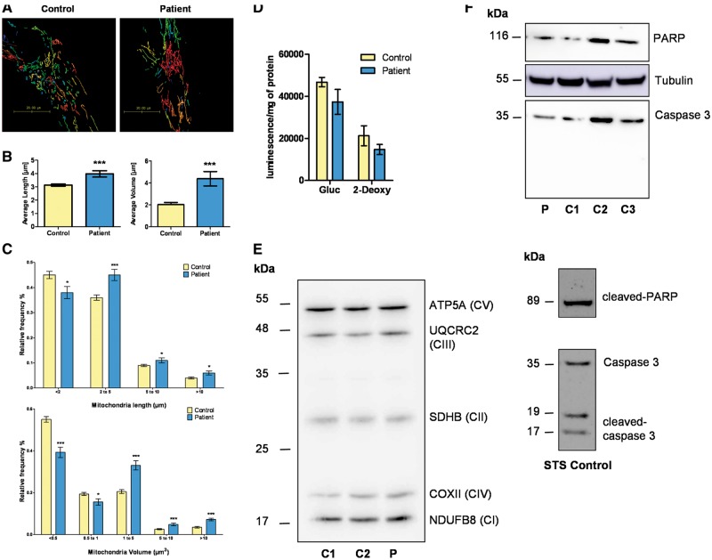
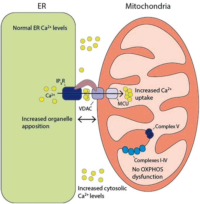
Wolfram syndrome (WS) is a progressive neurodegenerative disease characterized by early-onset optic atrophy and diabetes mellitus, which can be associated with more extensive central nervous system and endocrine complications. The majority of patients harbour pathogenic WFS1 mutations, but recessive mutations in a second gene, CISD2, have been described in a small number of families with Wolfram syndrome type 2 (WFS2). The defining diagnostic criteria for WFS2 also consist of optic atrophy and diabetes mellitus, but unlike WFS1, this phenotypic subgroup has been associated with peptic ulcer disease and an increased bleeding tendency. Here, we report on a novel homozygous CISD2 mutation (c.215A > G; p.Asn72Ser) in a Moroccan patient with an overlapping phenotype suggesting that Wolfram syndrome type 1 and type 2 form a continuous clinical spectrum with genetic heterogeneity. The present study provides strong evidence that this particular CISD2 mutation disturbs cellular Ca2+ homeostasis with enhanced Ca2+ flux from the ER to mitochondria and cytosolic Ca2+ abnormalities in patient-derived fibroblasts. This Ca2+ dysregulation was associated with increased ER-mitochondria contact, a swollen ER lumen and a hyperfused mitochondrial network in the absence of overt ER stress. Although there was no marked alteration in mitochondrial bioenergetics under basal conditions, culture of patient-derived fibroblasts in glucose-free galactose medium revealed a respiratory chain defect in complexes I and II, and a trend towards decreased ATP levels. Our results provide important novel insight into the potential disease mechanisms underlying the neurodegenerative consequences of CISD2 mutations and the subsequent development of multisystemic disease.


Figures

Figure 1
Figure 1
Brain magnetic resonance imaging of the proband. (A) Axial T1. Marked brainstem and cerebellar atrophy. (B) Axial T2 FLAIR. Cortical and subcortical atrophy with ventricular enlargement. (C) Sagittal T1. Brainstem and cerebellar atrophy, and lack of the normal posterior hypophysis hyperintense signal (arrow).
Figure 2
Figure 2
Identification of a novel CISD2 variant (c.215A > G; p.Asn72Ser) in the proband. (A) Family pedigree with the solid symbols representing clinically affected individuals. (B) Sequence chromatograms of the proband, his mother and his healthy sister. (C) Cross-species protein conservation of CISD2 in the region of the altered Asparagine amino acid at position 72. (D) In silico analysis of the CISD2 homodimeric model. The individual monomers have been coloured in pink and light blue. The domain topology of the dimeric CISD2 complex consists of a six-stranded β sandwich, which forms an intertwined β cap, and a larger cluster-binding domain carrying two 2Fe-2S clusters (one per protomer) that have been highlighted as grey spheres. The Asn72 residue (shown in red) is localized in a random coil region of the cluster-binding domain and it forms potential hydrogen bonds with the neighbouring amino acids Leu73, Ile75, Asp123 and Asn124, which are represented in light blue, green, yellow and dark blue, respectively. Our in silico modeling indicates that the p.Asn72Ser change alters the interactions necessary for the stabilization of the cluster-binding domains and this conformational change is predicted to affect the redox and functional properties of the CISD2 protein.
Figure 3
Figure 3
Absence of CISD2 RNA mis-splicing and normal CISD2 protein levels in the patient’s fibroblasts. (A) A 886bp amplicon was obtained from cDNAs using primers in the 5’-UTR and 3’-UTR which allow the amplification of the 3 CISD2 exons. M: molecular weight marker, P: patient, C: control; (-) negative controlc. (B) Western blot analysis with an anti-CISD2 antibody in fibroblasts from patient (P) and 3 control individuals (C1-C3).
Figure 4
Figure 4
Patient’s fibroblasts showed disturbed cellular Ca2+ homeostasis. (A) Imaging of cellular Ca2+ flux with Ca2+ sensitive fluorescent dyes. Representative images demonstrating analysis of mitochondrial Ca2+ with the Rhod-2-AM (red) dye (a, b), and cytosolic Ca2+ with the Fluo-4-AM (green) dye (c, d), before and after histamine stimulation. The relevant regions of interest were analysed using the Fiji open access software. (B) Representative traces of mitochondrial Ca2+ transients. (C) Analysis of average peak mitochondrial Ca2+ following histamine stimulation. F indicates fluorescence and F0 indicates basal fluorescence. Data are mean ± SEM (Control n = 35, patient n =15); * P ≤0.05; two-tailed unpaired t test. (D) Representative traces of cytosolic Ca2+ transients. (E) Analysis of average peak cytosolic Ca2+ following histamine stimulation. Data are mean ± SEM (Control n = 24, patient n = 14); * P ≤ 0.05; two-tailed unpaired t test. (F) Imaging with the D1ER probe. Representative images of the patient’s fibroblasts expressing D1ER: CFP signal (a) and YFP signal (b). The reticular pattern is indicative of ER localization. Regions of interest were analysed using the Fiji open access software. (G) Increased basal cytosolic [Ca2+] in the patient’s fibroblasts compared with controls. Data are mean ± SEM (n = 3); **P ≤0.01; two-tailed unpaired t test. (H) Analysis of peak cytosolic [Ca2+] levels after thapsigargin-induced ER Ca2+ depletion. Data are mean ± SEM (n = 3). (I) Comparison of thapsigargin (Tg)-sensitive Ca2+ stores in the patient’s fibroblasts and controls. Data are mean ± SEM (n = 3).
Figure 5
Figure 5
Increased ER-mitochondrial contacts observed in the patient’s fibroblasts. (A) Ultrastructural electron micrographs of fibroblasts from the patient (a-b) and a control individual (c-d). The contact sites between the ER and mitochondria have been indicated with arrows. The swollen ER lumen found in the patient’s cells has been highlighted with white asterisks. A higher magnification view of the typical mitochondrial morphology found in the patient’s fibroblasts has also been provided (inset of panel a). Scale bar = 1μm. (B) Quantification of the number of ER-mitochondria contacts expressed in mm2, as a percentage of mitochondria with ER contact sites, and as a percentage of the total mitochondria length adjacent to the ER normalized by mitochondrial perimeter. Data are mean ± SEM (Control n = 10, patient n = 10); ** P ≤0.01; * P ≤0.05; two-tailed unpaired t test. (C) Representative images of the ER and mitochondrial network (upper panels). Colocalisation analysis was performed using the Huygens Essential Analyzer and expressed as Manders’ coefficient M1 (ER/Mitochondrial (Mito) colocalisation) and M2 (Mito/ER colocalisation) to compare the patient’s fibroblasts with controls (lower panels). Data are mean ± SEM (Control n = 56, patient n = 26); *** P ≤0.001; * P ≤0.05; two-tailed unpaired t test. (D) qRT-PCR analysis of the ER stress markers BIP, CHOP and total XBP1. Data are mean ± SEM (n = 4); *** P ≤0.001; * P ≤0.05; two-tailed unpaired t test.
Figure 6
Figure 6
No evidence of major OXPHOS dysfunction or increased apoptosis in the patient’s fibroblasts. (A) Representative examples of the 3D-reconstructed mitochondrial networks in the patient’s fibroblasts and controls. Huygens Object Analyzer was used to determine the length and volume of each mitochondrial fragment. (B) Analysis of average mitochondrial fragment length (left panel) and volume (right panel). Data are mean ± SEM (Control n = 56, patient’s cells n = 26); *** P ≤0.001; two-tailed unpaired t test. (C) Distribution of mitochondrial fragment length (upper panel) and volume (lower panel). Data are mean ± SEM (n > 25); *** P ≤0.0001; * P ≤ 0.05; two-tailed unpaired t test. (D) ATP content assessed under two conditions: glucose (Gluc) and 2-deoxy-D-glucose (2-Deoxy). Data are mean ± SEM (n = 3); two-tailed unpaired t test. (E) Expression level of OXPHOS proteins. Representative western blot of ATP5A, UQCRC2, SDHB, COX II and NDUFB8 proteins performed with fibroblast lysates obtained from two control individuals (C1, C2) and the patient (P). CI-CV; complex I-V. (F) Representative western blots of caspase 3 and PARP in untreated controls (C1-C3) and the patient’s fibroblasts (P) (upper panel). The caspase 3 and PARP antibodies had previously been validated on an STS (staurosporin)-treated control fibroblast (lower panel).
Figure 7
Figure 7
Schematic model highlighting the deleterious consequences of the CISD2 (c.215A > G; p.Asn72Ser) mutation. This mutation was associated with disturbed Ca2+ homeostasis characterised by enhanced ER-mitochondrial Ca2+ flux and increased cytosolic [Ca2+]. There was increased ER-mitochondrial interorganellar contact in the absence of any major disturbance in OXPHOS dysfunction.

Similar articles

Cited by

References

    1. Barrett T.G., Bundey S.E., Macleod A.F. (1995) Neurodegeneration and diabetes: UK nationwide study of Wolfram (DIDMOAD) syndrome. Lancet Lond. Engl., 346, 1458–1463. - PubMed
    1. Khanim F., Kirk J., Latif F., Barrett T.G. (2001) WFS1/wolframin mutations, Wolfram syndrome, and associated diseases. Hum. Mutat., 17, 357–367. - PubMed
    1. Amr S., Heisey C., Zhang M., Xia X.-J., Shows K.H., Ajlouni K., Pandya A., Satin L.S., El-Shanti H., Shiang R. (2007) A homozygous mutation in a novel zinc-finger protein, ERIS, is responsible for Wolfram syndrome 2. Am. J. Hum. Genet., 81, 673–683. - PMC - PubMed
    1. Mozzillo E., Delvecchio M., Carella M., Grandone E., Palumbo P., Salina A., Aloi C., Buono P., Izzo A., D’Annunzio G., et al. (2014) A novel CISD2 intragenic deletion, optic neuropathy and platelet aggregation defect in Wolfram syndrome type 2. BMC Med. Genet., 15, 88.. - PMC - PubMed
    1. Rondinelli M., Novara F., Calcaterra V., Zuffardi O., Genovese S. (2015) Wolfram syndrome 2: a novel CISD2 mutation identified in Italian siblings. Acta Diabetol., 52, 175–178. - PubMed

Publication types

Supplementary concepts