

Multifunctional Inorganic Nanoparticles: Recent Progress in Thermal Therapy and Imaging
- PMID: 28335204
- PMCID: PMC5302572
- DOI: 10.3390/nano6040076
Multifunctional Inorganic Nanoparticles: Recent Progress in Thermal Therapy and Imaging
Abstract
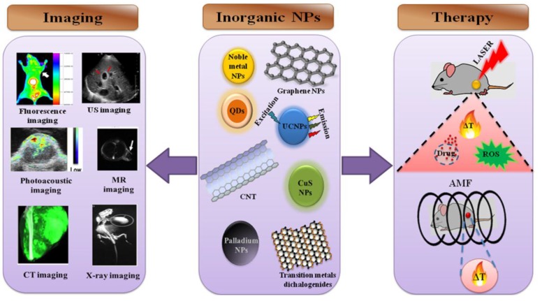
Nanotechnology has enabled the development of many alternative anti-cancer approaches, such as thermal therapies, which cause minimal damage to healthy cells. Current challenges in cancer treatment are the identification of the diseased area and its efficient treatment without generating many side effects. Image-guided therapies can be a useful tool to diagnose and treat the diseased tissue and they offer therapy and imaging using a single nanostructure. The present review mainly focuses on recent advances in the field of thermal therapy and imaging integrated with multifunctional inorganic nanoparticles. The main heating sources for heat-induced therapies are the surface plasmon resonance (SPR) in the near infrared region and alternating magnetic fields (AMFs). The different families of inorganic nanoparticles employed for SPR- and AMF-based thermal therapies and imaging are described. Furthermore, inorganic nanomaterials developed for multimodal therapies with different and multi-imaging modalities are presented in detail. Finally, relevant clinical perspectives and the future scope of inorganic nanoparticles in image-guided therapies are discussed.
Keywords: alternate magnetic field; image-guided therapy; imaging; inorganic nanoparticles; photothermal therapy; surface plasmon resonance.
Conflict of interest statement
The authors have no other relevant affiliations or financial involvement with any organization or entity with a financial interest in or financial conflict with the subject matter or materials discussed in the manuscript. This includes employment, consultancies, stock ownership or options, expert testimony, grants or patents received or pending, or royalties.
Figures

Similar articles
-
Current Challenges in Image-Guided Magnetic Hyperthermia Therapy for Liver Cancer.Nanomaterials (Basel). 2022 Aug 12;12(16):2768. doi: 10.3390/nano12162768. Nanomaterials (Basel). 2022. PMID: 36014633 Free PMC article. Review.
-
Near-infrared inorganic nanomaterial-based nanosystems for photothermal therapy.Nanoscale. 2021 May 20;13(19):8751-8772. doi: 10.1039/d1nr00323b. Nanoscale. 2021. PMID: 33973616 Review.
-
Functionalized Magnetic Nanoparticles for Alternating Magnetic Field- or Near Infrared Light-Induced Cancer Therapies.Micromachines (Basel). 2022 Aug 8;13(8):1279. doi: 10.3390/mi13081279. Micromachines (Basel). 2022. PMID: 36014201 Free PMC article. Review.
-
Nanotechnology: an evidence-based analysis.Ont Health Technol Assess Ser. 2006;6(19):1-43. Epub 2006 Nov 1. Ont Health Technol Assess Ser. 2006. PMID: 23074489 Free PMC article.
-
Magnetic Heating Stimulated Cargo Release with Dose Control using Multifunctional MR and Thermosensitive Liposome.Nanotheranostics. 2019 Apr 19;3(2):166-178. doi: 10.7150/ntno.31164. eCollection 2019. Nanotheranostics. 2019. PMID: 31183312 Free PMC article.
Cited by
-
Role of Immunosuppressive Microenvironment in Acquiring Immunotolerance Post-Photothermal Therapy.J Korean Med Sci. 2019 Nov 18;34(44):e272. doi: 10.3346/jkms.2019.34.e272. J Korean Med Sci. 2019. PMID: 31726492 Free PMC article.
-
A Step Forward for the Treatment of Localized Prostate Cancer Using Gold Nanoparticles Combined with Laser Irradiation.Int J Mol Sci. 2024 Apr 19;25(8):4488. doi: 10.3390/ijms25084488. Int J Mol Sci. 2024. PMID: 38674073 Free PMC article.
-
The Application of Nanoparticles in Diagnosis and Treatment of Kidney Diseases.Int J Mol Sci. 2021 Dec 23;23(1):131. doi: 10.3390/ijms23010131. Int J Mol Sci. 2021. PMID: 35008556 Free PMC article. Review.
-
Europium-phenolic network coated BaGdF5 nanocomposites for tri-modal computed tomography/magnetic resonance/luminescence imaging.J Mater Sci Mater Med. 2017 May;28(5):74. doi: 10.1007/s10856-017-5888-5. Epub 2017 Mar 30. J Mater Sci Mater Med. 2017. PMID: 28361281
-
Recent Advances in Copper-Based Organic Complexes and Nanoparticles for Tumor Theranostics.Molecules. 2022 Oct 19;27(20):7066. doi: 10.3390/molecules27207066. Molecules. 2022. PMID: 36296659 Free PMC article. Review.
References
Publication types
LinkOut - more resources
Full Text Sources
Other Literature Sources
