Dye-Doped Fluorescent Silica Nanoparticles for Live Cell and In Vivo Bioimaging
- PMID: 28335209
- PMCID: PMC5302498
- DOI: 10.3390/nano6050081
Dye-Doped Fluorescent Silica Nanoparticles for Live Cell and In Vivo Bioimaging
Abstract
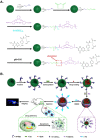
The need for novel design strategies for fluorescent nanomaterials to improve our understanding of biological activities at the molecular level is increasing rapidly. Dye-doped fluorescent silica nanoparticles (SiNPs) emerge with great potential for developing fluorescence imaging techniques as a novel and ideal platform for the monitoring of living cells and the whole body. Organic dye-containing fluorescent SiNPs exhibit many advantages: they have excellent biocompatibility, are non-toxic, highly hydrophilic, optically transparent, size-tunable and easily modified with various biomolecules. The outer silica shell matrix protects fluorophores from outside chemical reaction factors and provides a hydrophilic shell for the insoluble nanoparticles, which enhances the photo-stability and biocompatibility of the organic fluorescent dyes. Here, we give a summary of the synthesis, characteristics and applications of fluorescent SiNPs for non-invasive fluorescence bioimaging in live cells and in vivo. Additionally, the challenges and perspectives of SiNPs are also discussed. We prospect that the further development of these nanoparticles will lead to an exciting breakthrough in the understanding of biological processes.
Keywords: bioimaging; in vivo; organic fluorescent dye; silica nanoparticle.
Conflict of interest statement
The authors declare no conflict of interest.
Figures


Similar articles
-
Functionalized silica nanoparticles: a platform for fluorescence imaging at the cell and small animal levels.Acc Chem Res. 2013 Jul 16;46(7):1367-76. doi: 10.1021/ar3001525. Epub 2013 Mar 14. Acc Chem Res. 2013. PMID: 23489227 Review.
-
A two-photon fluorescence silica nanoparticle-based FRET nanoprobe platform for effective ratiometric bioimaging of intracellular endogenous adenosine triphosphate.Analyst. 2021 Jul 26;146(15):4945-4953. doi: 10.1039/d1an00419k. Analyst. 2021. PMID: 34259245
-
A novel fluorescent label based on organic dye-doped silica nanoparticles for HepG liver cancer cell recognition.J Nanosci Nanotechnol. 2004 Jul;4(6):585-9. doi: 10.1166/jnn.2004.011. J Nanosci Nanotechnol. 2004. PMID: 15518391
-
Fluorescence resonance energy transfer mediated large Stokes shifting near-infrared fluorescent silica nanoparticles for in vivo small-animal imaging.Anal Chem. 2012 Nov 6;84(21):9056-64. doi: 10.1021/ac301461s. Epub 2012 Oct 24. Anal Chem. 2012. PMID: 23017033
-
Silicon nanomaterials platform for bioimaging, biosensing, and cancer therapy.Acc Chem Res. 2014 Feb 18;47(2):612-23. doi: 10.1021/ar400221g. Epub 2014 Jan 7. Acc Chem Res. 2014. PMID: 24397270 Review.
Cited by
-
Nanomaterials for Biosensing Lipopolysaccharide.Biosensors (Basel). 2019 Dec 21;10(1):2. doi: 10.3390/bios10010002. Biosensors (Basel). 2019. PMID: 31877825 Free PMC article. Review.
-
An insight into the in vivo imaging potential of curcumin analogues as fluorescence probes.Asian J Pharm Sci. 2021 Jul;16(4):419-431. doi: 10.1016/j.ajps.2020.11.003. Epub 2020 Dec 5. Asian J Pharm Sci. 2021. PMID: 34703492 Free PMC article. Review.
-
Optimizing the performance of silica nanoparticles functionalized with a near-infrared fluorescent dye for bioimaging applications.Nanotechnology. 2024 May 9;35(30):10.1088/1361-6528/ad3fc5. doi: 10.1088/1361-6528/ad3fc5. Nanotechnology. 2024. PMID: 38631329 Free PMC article.
-
Tandem Dye-Doped Nanoparticles for NIR Imaging via Cerenkov Resonance Energy Transfer.Front Chem. 2020 Feb 27;8:71. doi: 10.3389/fchem.2020.00071. eCollection 2020. Front Chem. 2020. PMID: 32175305 Free PMC article.
-
Functionalized Fluorescent Silica Nanoparticles for Bioimaging of Cancer Cells.Sensors (Basel). 2020 Sep 29;20(19):5590. doi: 10.3390/s20195590. Sensors (Basel). 2020. PMID: 33003513 Free PMC article.
References
-
- Perkel J.M. Cell signaling: In vivo veritas. Science. 2007;316:1763–1768. doi: 10.1126/science.316.5832.1763. - DOI
-
- Koo H., Huh M.S., Ryu J.H., Lee D.-E., Sun I.-C., Choi K., Kim K., Kwon I.C. Nanoprobes for biomedical imaging in living systems. Nano Today. 2011;6:204–220. doi: 10.1016/j.nantod.2011.02.007. - DOI
Publication types
LinkOut - more resources
Full Text Sources
Other Literature Sources

