. 2016 May 9;6(5):87.
doi: 10.3390/nano6050087.

Rapamycin Loaded Solid Lipid Nanoparticles as a New Tool to Deliver mTOR Inhibitors: Formulation and in Vitro Characterization

Affiliations

Rapamycin Loaded Solid Lipid Nanoparticles as a New Tool to Deliver mTOR Inhibitors: Formulation and in Vitro Characterization

Alice Polchi et al. Nanomaterials (Basel). .

Abstract

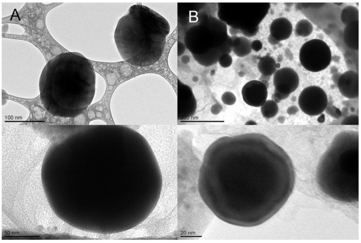
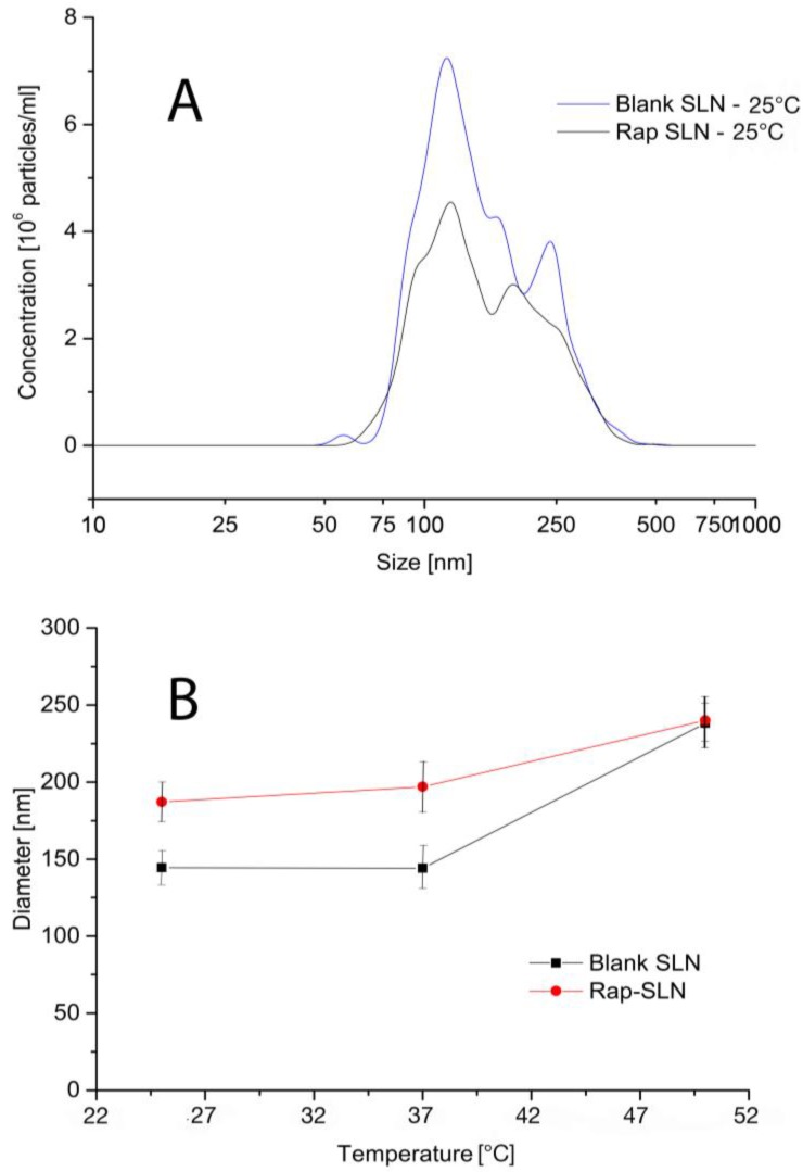
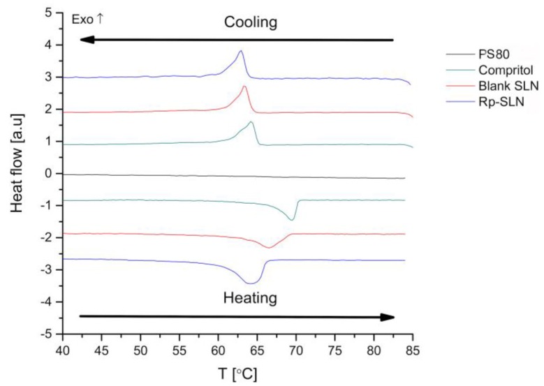
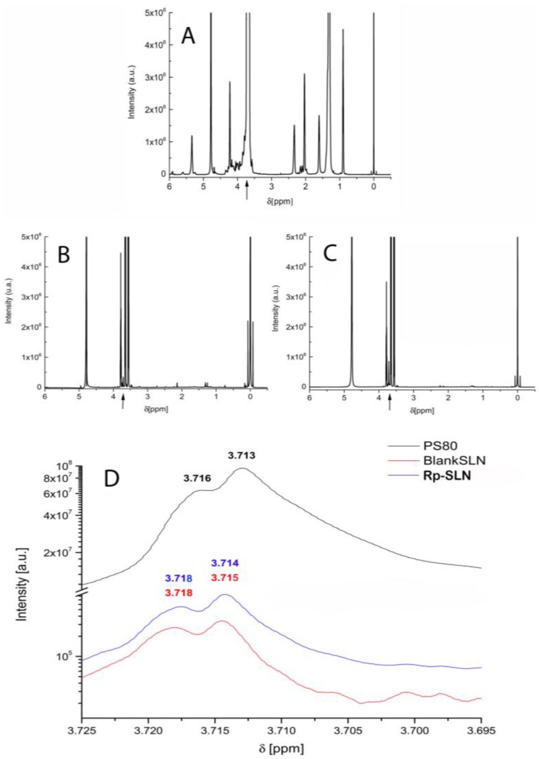
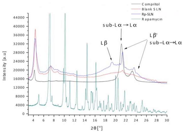
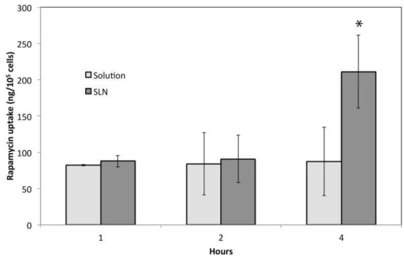
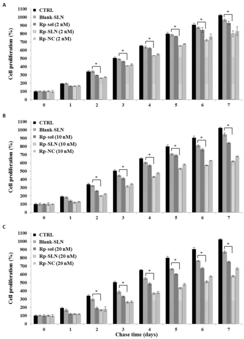
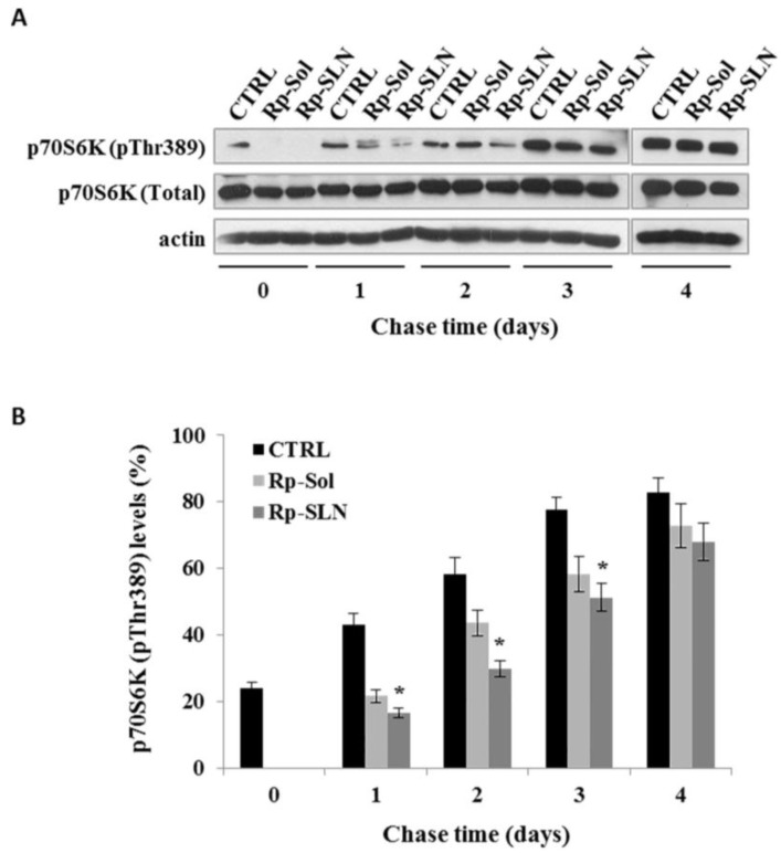
Recently, the use of mammalian target of rapamycin (mTOR) inhibitors, in particular rapamycin (Rp), has been suggested to improve the treatment of neurodegenerative diseases. However, as Rp is a strong immunosuppressant, specific delivery to the brain has been postulated to avoid systemic exposure. In this work, we fabricated new Rp loaded solid lipid nanoparticles (Rp-SLN) stabilized with polysorbate 80 (PS80), comparing two different methods and lipids. The formulations were characterized by differential scanning calorimetry (DSC), nuclear magnetic resonance (NMR), wide angle X-ray scattering (WAXS), cryo-transmission electron microscopy (cryo-TEM), dynamic light scattering (DLS) and particle tracking. In vitro release and short-term stability were assessed. Biological behavior of Rp-SLN was tested in SH-SY5Y neuroblastoma cells. The inhibition of mTOR complex 1 (mTORC1) was evaluated over time by a pulse-chase study compared to free Rp and Rp nanocrystals. Compritol Rp-SLN resulted more stable and possessing proper size and surface properties with respect to cetyl palmitate Rp-SLN. Rapamycin was entrapped in an amorphous form in the solid lipid matrix that showed partial crystallinity with stable Lβ, sub-Lα and Lβ' arrangements. PS80 was stably anchored on particle surface. No drug release was observed over 24 h and Rp-SLN had a higher cell uptake and a more sustained effect over a week. The mTORC1 inhibition was higher with Rp-SLN. Overall, compritol Rp-SLN show suitable characteristics and stability to be considered for further investigation as Rp brain delivery system.

Keywords: SH-SY5Y neuroblastoma cells; drug delivery; formulation; rapamycin; solid lipid nanoparticles.


Conflict of interest statement

The authors declare no conflicts of interest. The founding sponsor had no role in the design of the study; in the collection, analyses, or interpretation of data; in the writing of the manuscript, and in the decision to publish the results.

Figures

Figure 1
Figure 1
Hydrodynamic size and polydispersity change over time for Rp loaded compritol and cetyl palmitate SLN.
Figure 2
Figure 2
Cryo-transmission electron microscopy (cryo-TEM) images of blank (A) and Rp-SLN (B). Magnification 180,000× and 200,000×.
Figure 3
Figure 3
Particle tracking: dimensional profiles of blank and Rp-SLN at 25 °C (A) and an effect of temperature on the mean MHD of blank and Rp-SLN (B).
Figure 4
Figure 4
Heating and cooling ramps of blank and Rp-SLN compared to bulk compritol and PS80.
Figure 5
Figure 5
Proton nuclear magnetic resonance spectroscopy (1H NMR) spectra of (A) PS80; (B) blank and (C) Rp-SLN all prepared in D2O, were submitted to an external magnetic field of 18.8 T and 1H resonance frequency of 800 MHz. The arrows indicate the area magnified in (D) corresponding to the PS80-derived oxyethylene moiety signals (about δ = 3.7) to highlight the slight shift occurring in SLN compared to pure PS80.
Figure 6
Figure 6
WAXS profiles of pure Rp, compritol and blank and Rp-SLN. Arrows indicate the signals corresponding to the polymorphs observed for blank, Rp-SLN and bulk lipid.
Figure 7
Figure 7
Cell uptake of SLN. Amount of Rp taken up by SH-SY5Y cells after 1, 2 and 4 h after the treatment with 200 nM Rp-SLN and Rp solution. * p < 0.001.
Figure 8
Figure 8
Cell uptake of fluorescent-SLN. Fluorescence microscopy images of SH-SY5Y cells were taken 1(A); 2 (B) and 4 h (C) after treatment with 500 nM DiQ-tagged Rp-SLN DiQ (red) and after staining of lysosomes with fluorescein isothiocyanate dextran (green). Nuclei were stained with 4',6-diamidino-2-phenylindole (DAPI). Magnification: 60×.
Figure 9
Figure 9
Effect of the Rp treatments on cell proliferation. The Rp effect was evaluated in SH-SY5Y cells by pulse-chase experiments. Cells were seeded in a 96-well plate, incubated overnight at 37 °C and treated for 4 h with Rp solution (Rp-sol), Rp-nanocrystals (Rp-NC) and Rp-SLN at the concentration of 2, 10 and 20 nM (Pulse), panel (AC), respectively. The cells were also treated with blank SLN as control. The cell proliferation was evaluated daily by using 3-[4,5-dimethylthiazol-2-yl]-2,5-diphenyltetrazolium bromide (MTT) assay. Control (CTRL): untreated cells. Values are the mean ± S.D. of three independent experiments. * p < 0.01 (Rp-SLN vs. blank SLN cells) according to unpaired two-tailed Student’s t-test.
Figure 10
Figure 10
Effect of Rp-SLN on mammalian target of rapamycin (mTOR) activity. The SH-SY5Y cells were seeded in a 6-well plate, incubated overnight at 37 °C and then treated for 4 h with 20 nM of Rp-sol or Rp-SLN (Pulse); untreated cells were considered as control (CTRL). (A) Cells were recovered after 0, 1, 2, 3 and 4 days (chase) and the immunoblotting analysis was performed for phospho-p70S6K (pThr389), p70S6K (Total) and actin. Representative Western blotting of three independent experiments is reported; (B) densitometric analysis of the immunoblot represents the percentage of the ratio between phospho-p70S6K (pThr389) with respect to p70S6K. Values are the mean ± S.D. of three independent experiments. * p < 0.01 (Rp-SLN vs. CTRL cells) according to unpaired two tailed Student’s t-test.

Similar articles

Cited by

References

    1. Balmayor E.R., Azevedo H.S., Reis R.L. Controlled delivery systems: From pharmaceuticals to cells and genes. Pharm. Res. 2011;28:1241–1258. doi: 10.1007/s11095-011-0392-y. - DOI - PubMed
    1. Blasi P., Giovagnoli S., Schoubben A., Ricci M., Rossi C. Solid lipid nanoparticles for targeted brain drug delivery. Adv. Drug Deliv. Rev. 2007;59:454–477. doi: 10.1016/j.addr.2007.04.011. - DOI - PubMed
    1. Martins S., Costa-Lima S., Carneiro T., Cordeiro-da-Silva A., Souto E.B., Ferreira D.C. Solid lipid nanoparticles as intracellular drug transporters: An investigation of the uptake mechanism and pathway. Int. J. Pharm. 2012;430:216–227. doi: 10.1016/j.ijpharm.2012.03.032. - DOI - PubMed
    1. Gastaldi L., Battaglia L., Peira E., Chirio D., Muntoni E., Solazzi I., Gallarate M., Dosio F. Solid lipid nanoparticles as vehicles of drugs to the brain: Current state of the art. Eur. J. Pharm. Biopharm. 2014;87:433–444. doi: 10.1016/j.ejpb.2014.05.004. - DOI - PubMed
    1. Göppert T.M., Müller R.H. Polysorbate-stabilized solid lipid nanoparticles as colloidal carriers for intravenous targeting of drugs to the brain: Comparison of plasma protein adsorption patterns. J. Drug Target. 2005;13:179–187. doi: 10.1080/10611860500071292. - DOI - PubMed

LinkOut - more resources