Review
. 2016 Jul 19;6(7):133.
doi: 10.3390/nano6070133.

Nanomaterials for Cardiac Myocyte Tissue Engineering

Affiliations
Review

Nanomaterials for Cardiac Myocyte Tissue Engineering

Rodolfo Amezcua et al. Nanomaterials (Basel). .

Abstract

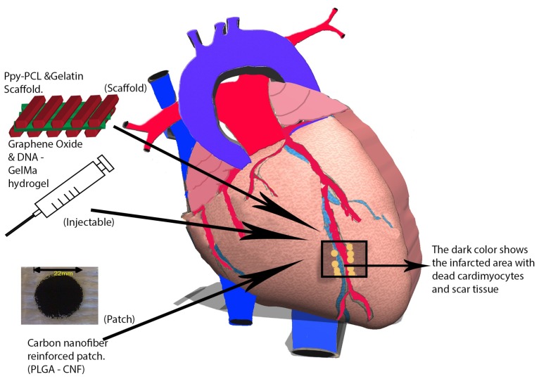
Since their synthesizing introduction to the research community, nanomaterials have infiltrated almost every corner of science and engineering. Over the last decade, one such field has begun to look at using nanomaterials for beneficial applications in tissue engineering, specifically, cardiac tissue engineering. During a myocardial infarction, part of the cardiac muscle, or myocardium, is deprived of blood. Therefore, the lack of oxygen destroys cardiomyocytes, leaving dead tissue and possibly resulting in the development of arrhythmia, ventricular remodeling, and eventual heart failure. Scarred cardiac muscle results in heart failure for millions of heart attack survivors worldwide. Modern cardiac tissue engineering research has developed nanomaterial applications to combat heart failure, preserve normal heart tissue, and grow healthy myocardium around the infarcted area. This review will discuss the recent progress of nanomaterials for cardiovascular tissue engineering applications through three main nanomaterial approaches: scaffold designs, patches, and injectable materials.

Keywords: cardiac infarction; injectable; nanomaterials; patch; scaffold; tissue engineering.


Figures

Figure 1
Figure 1
During a myocardial infarction, blood flow is deprived to the myocardium, which, in turn, injures the tissues downstream of the obstruction. As a result, cardiomyocytes are deprived of oxygen and eventually perish. Next, myofibroblasts migrate towards this infarct area and ultimately create scar tissue that reduces the heart’s contractile ability and weakens it. The weakened area is unable to withstand the pressure and volume load on the heart, thereby causing left ventricular dilation. Over time, this scar tissue causes ongoing remodeling, and the heart becomes more spherical, thus impairing systolic and diastolic function. To combat the progress of cardiovascular disease (CVD) and ventricular remodeling, modern research has deployed the use of nanomaterials through the use of scaffolds, patches, and injectable materials for regenerating the heart tissue.
Figure 2
Figure 2
Researchers have exploited nanomaterial characteristics for biomaterial applications by increasing its surface-area ratio. This augmentation in surface-area ratio can enhance protein absorption, thus increasing the possibility of cell recruitment and attachment.
Figure 3
Figure 3
Two novel studies in cardiac tissue engineering: (a) electrospun polycaprolactone (PCL) nanofibrous mesh stretched across a wire ring used to create a passive load exerted by the wire allowing for cultured cardiomyocytes (CMs) to reach appropriate maturity prior to implantation [52]; (b) nanoelectronic scaffold (blue) constructed from silicon nanowire field effect transistors capable of real-time monitoring of local electrical activity within the extracellular matrix (ECM) (green) [55].
Figure 4
Figure 4
Cardiovascular patches embedded with nanomaterials play a key role in promoting stem cell growth around an infarct area of the myocardium: (a) the use of poly(lactic-co-glycolic) acid (PLGA) embedded with carbon nanofibers (CNFs) to generate a carbon nanofiber reinforced patch; (b) scanning electron micrograph of carbon nanofiber reinforced patch showing CNFs embedded in PLGA (at 10 K magnification); (c) atomic force micrograph of a carbon nanofiber reinforced patch depicting the topography of the patch; (d) using the data from atomic force micrographs to calculate the increase in surface area as one increases CNF concentration vs. protein adsorption, one can see that using CNFs can increase protein adsorption of a carbon nanofiber reinforced patch, thus increasing the possibility of cell recruitment and growth.
Figure 5
Figure 5
Following cardiac ischemic injury, lack of blood supply results in cardiomyocyte apoptosis (brown). After an inflammatory response, myofibroblasts and macrophages (green) migrate towards the injured area leading to fibrosis thus creating scar tissue (white). When scaffolds are applied to the injured area (in this case by injection via syringe (clear)), exploiting the superior material properties of nanomaterials such as gold nanoparticles (yellow) have demonstrated reduced scarring and often slowed left ventricular remodeling. The (pink) cardiomyocytes in this figure are shown to regenerate along the site of scaffold injection.

References

    1. Mozaffarian D., Benjamin E.J., Go A.S., Arnett D.K., Blaha M.J., Cushman M., Das S.R., de Ferranti S., Despres J.P., Fullerton H.J., et al. Heart Disease and Stroke Statistics-2016 Update: A Report From the American Heart Association. Circulation. 2016;133:e38–e360. doi: 10.1161/CIR.0000000000000350. - DOI - PubMed
    1. Thygesen K., Alpert J.S., White H.D. Universal definition of myocardial infarction. J. Am. Coll. Cardiol. 2007;50:2173–2195. doi: 10.1016/j.jacc.2007.09.011. - DOI - PubMed
    1. Woodcock E.A., Matkovich S.J. Cardiomyocytes structure, function and associated pathologies. Int. J. Biochem. Cell Biol. 2005;37:1746–1751. doi: 10.1016/j.biocel.2005.04.011. - DOI - PubMed
    1. Vunjak-Novakovic G., Tandon N., Godier A., Maidhof R., Marsano A., Martens T.P., Radisic M. Challenges in cardiac tissue engineering. Tissue Eng. Part B Rev. 2009;16:169–187. doi: 10.1089/ten.teb.2009.0352. - DOI - PMC - PubMed
    1. Rodriguez L.E., Suarez E.E., Loebe M., Bruckner B.A. Ventricular assist devices (VAD) therapy: New technology, new hope? Methodist DeBakey Cardiovasc. J. 2013;9:32–37. doi: 10.14797/mdcj-9-1-32. - DOI - PMC - PubMed

LinkOut - more resources