. 2016 Aug 18;6(8):149.
doi: 10.3390/nano6080149.

Receptor-Meditated Endocytosis by Hyaluronic Acid@Superparamagnetic Nanovetor for Targeting of CD44-Overexpressing Tumor Cells

Affiliations

Receptor-Meditated Endocytosis by Hyaluronic Acid@Superparamagnetic Nanovetor for Targeting of CD44-Overexpressing Tumor Cells

Kwang Sik Yu et al. Nanomaterials (Basel). .

Abstract

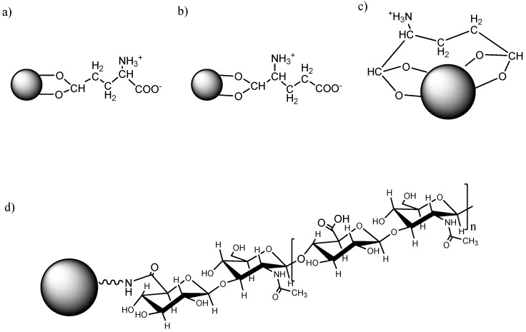
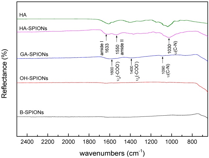
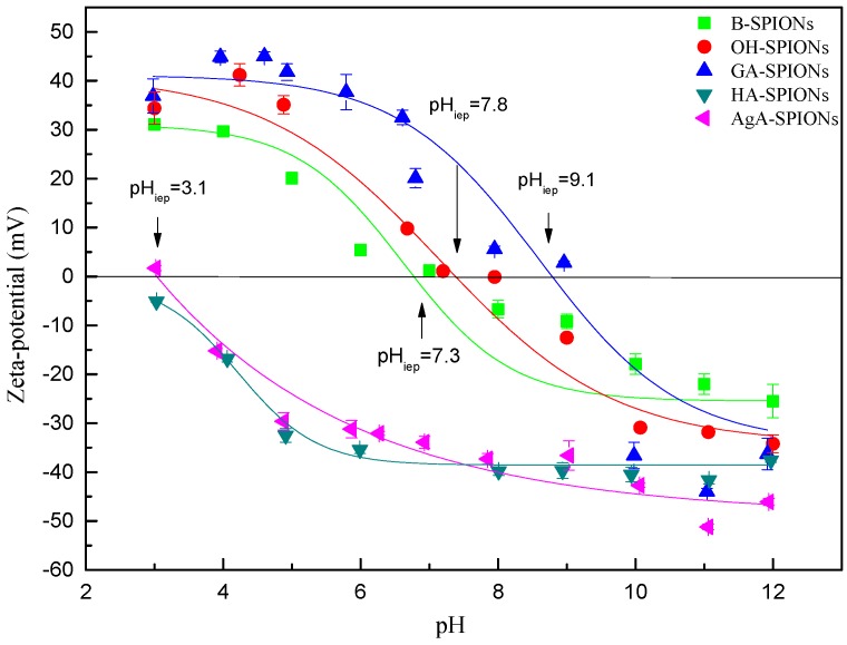
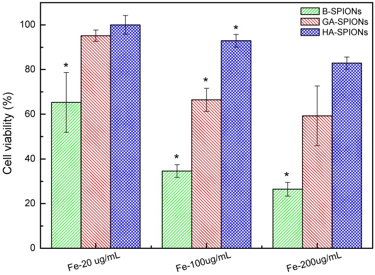
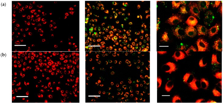
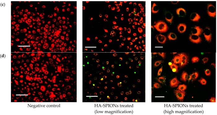
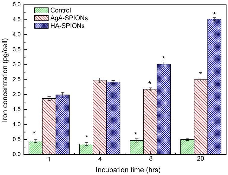
The present report proposes a more rational hyaluronic acid (HA) conjugation protocol that can be used to modify the surface of the superparamagnetic iron oxide nanoparticles (SPIONs) by covalently binding the targeting molecules (HA) with glutamic acid as a molecular linker on peripheral surface of SPIONs. The synthesis of HA-Glutamic Acid (GA)@SPIONs was included oxidization of nanoparticle's surface with H₂O₂ followed by activation of hydroxyl group and reacting glutamic acid as an intermediate molecule demonstrating transfection of lung cancer cells. Fourier transform infrared (FTIR) and zeta-potential studies confirmed the chemical bonding between amino acid linker and polysaccharides. 3-(4,5-dimethylthiazol-2-yl)-2,5-diphenyltetrazolium bromide (MTT) cytotoxicity assay showed that HA-SPIONs-treated cells remained 82.9% ± 2.7% alive at high particle dosage (200 µg/mL iron concentration), whereas GA-SPIONs and bare SPIONs (B-SPIONs) treated cells had only 59.3% ± 13.4% and 26.5% ± 3.1% survival rate at the same conditions, respectively. Confocal microscopy analysis showed increased cellular internalization of HA-SPIONs compared to non-interacting agarose coated SPIONs (AgA-SPIONs).

Keywords: CD44; glutamic acid; hyaluronan; receptor-meditated endocytosis (RME); superparamagnetic.


Figures

Figure 1
Figure 1
(ac) Three possible configurations of glutamic acid superparamagnetic iron oxide nanoparticles (GA-SPIONs); and (d) schematic illustration of hyaluronic acid superparamagnetic iron oxide nanoparticles (HA-SPIONs).
Figure 2
Figure 2
Transmission electron microscopy (TEM) images of: (a) bare SPIONs (B-SPIONs); (b) hydroxyl group activated SPION (OH-SPIONs); (c) GA-SPIONs; and (d) HA-SPIONs.
Figure 3
Figure 3
Fourier transform infrared spectra of B-SPIONs, OH-SPIONs, GA-SPIONs and HA-SPIONs in the wavenumber range of 2500 cm−1 to 650 cm−1.
Figure 4
Figure 4
Zeta-potential of B-SPIONs, OH-SPIONs, GA-SPIONs, HA-SPIONs and agarose coated SPIONs (AgA-SPIONs).
Figure 5
Figure 5
Histogram of cell viability after 24 h incubation with B-SPIONs (green), GA-SPIONs (red) and HA-SPIONs (blue) at 37 °C, 5% CO2. (* means the mean of the group is significantly different from the other two at the same Fe concentration at p = 0.05 level) (n ≥ 5).
Figure 6
Figure 6
Confocal microscopy images of A459 cells only as control (left column); A459 cells incubated with 20 µg/mL fluorescein isothiocyanate (FITC)-HA-SPIONs (green colored) (middle column) and enlarged imaged of A459 cells with FITC-HA-SPIONs (right column) for: (a) 6 h; (b) 8 h; (c) 12 h; and (d) 24 h. Cells were stained with red membrane dye PKH26 prior to FTIC-HA-SPIONs incubation at 37 °C, 5% CO2 (the scale bars of the left and middle column are 100 mm, and the scale bars of the right column are 20 mm).
Figure 6
Figure 6
Confocal microscopy images of A459 cells only as control (left column); A459 cells incubated with 20 µg/mL fluorescein isothiocyanate (FITC)-HA-SPIONs (green colored) (middle column) and enlarged imaged of A459 cells with FITC-HA-SPIONs (right column) for: (a) 6 h; (b) 8 h; (c) 12 h; and (d) 24 h. Cells were stained with red membrane dye PKH26 prior to FTIC-HA-SPIONs incubation at 37 °C, 5% CO2 (the scale bars of the left and middle column are 100 mm, and the scale bars of the right column are 20 mm).
Figure 7
Figure 7
Histogram of cellular iron uptake of A459 cells without any treatment (green) and those incubated with AgA-SPIONs (red) and HA-SPIONs (blue), cells with no particles treatment as control (green), at 1 h, 4 h, 8 h and 20 h incubation, at 37 °C, 5% CO2. (* means the mean of the group is significantly different from the other two groups at p = 0.05 level) (n ≥ 5).

Similar articles

Cited by

References

    1. Gorvin C.M., Wilmer M.J., Piret S.E., Harding B., van den Heuvel L.P., Wrong O., Jat P.S., Lippiat J.D., Levtchenko E.N., Thakker R.V. Receptor-mediated endocytosis and endosomal acidification is impaired in proximal tubule epithelial cells of dent disease patients. Proc. Natl. Acad. Sci. USA. 2013;110:7014–7019. doi: 10.1073/pnas.1302063110. - DOI - PMC - PubMed
    1. Ding H.-M., Ma Y.-Q. Role of physicochemical properties of coating ligands in receptor-mediated endocytosis of nanoparticles. Biomaterials. 2012;33:5798–5802. doi: 10.1016/j.biomaterials.2012.04.055. - DOI - PubMed
    1. Sun C., Sze R., Zhang M. Folic acid-peg conjugated superparamagnetic nanoparticles for targeted cellular uptake and detection by mri. J. Biomed. Mater. Res. 2006;78:550–557. doi: 10.1002/jbm.a.30781. - DOI - PubMed
    1. Saraswati T.E., Ogino A., Nagatsu M. Plasma-activated immobilization of biomolecules onto graphite-encapsulated magnetic nanoparticles. Carbon. 2012;50:1253–1261. doi: 10.1016/j.carbon.2011.10.044. - DOI
    1. Chen J.-T., Ahmed M., Liu Q., Narain R. Synthesis of cationic magnetic nanoparticles and evaluation of their gene delivery efficacy in hep g2 cells. J. Biomed. Mater. Res. 2012;100:2342–2347. doi: 10.1002/jbm.a.34176. - DOI - PubMed