. 2016 Dec 20;6(12):243.
doi: 10.3390/nano6120243.

Synthesis of Antifungal Agents from Xanthene and Thiazine Dyes and Analysis of Their Effects

Affiliations

Synthesis of Antifungal Agents from Xanthene and Thiazine Dyes and Analysis of Their Effects

Joo Ran Kim et al. Nanomaterials (Basel). .

Abstract

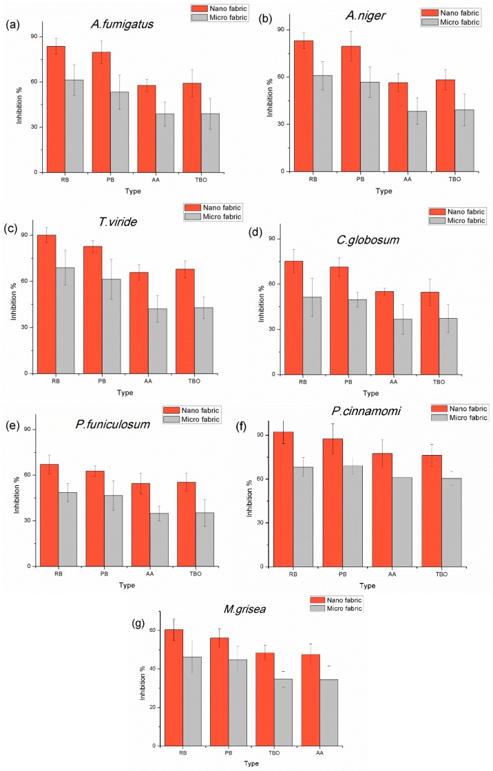
Indoor fungi growth is an increasing home health problem as our homes are more tightly sealed. One thing that limits durability of the antifungal agents is the scarcity of reactive sites on many surfaces to attach these agents. In order to increase graft yield of photosensitizers to the fabrics, poly(acrylic acid-co-styrene sulfonic acid-co-vinyl benzyl rose bengal or phloxine B) were polymerized and then grafted to electrospun fabrics. In an alternative process, azure A or toluidine blue O were grafted to poly(acrylic acid), which was subsequently grafted to nanofiber-based and microfiber-based fabrics. The fabrics grafted with photosensitizers induced antifungal effects on all seven types of fungi in the order of rose bengal > phloxine B > toluidine blue O > azure A, which follows the quantum yield production of singlet oxygen for these photoactive dyes. Their inhibition rates for inactivating fungal spores decreased in the order of P. cinnamomi, T. viride, A. niger, A. fumigatus, C. globosum, P. funiculosum, and M. grisea, which is associated with lipid composition in membrane and the morphology of fungal spores. The antifungal activity was also correlated with the surface area of fabric types which grafted the photosensitizer covalently on the surface as determined by the bound color strength.

Keywords: Aspergillus; Chaetomium; Magnaporthe; antifungal photosensitizer; nanofiber.


Conflict of interest statement

The authors declare no conflict of interest.

Figures

Figure 1
Figure 1
Nanofibers using electrospinning using roller collector, (a) nylon 6,6 fabric consisting of nanofibers with average diameter 505 nm (σ = 152.5 nm); and (b) melt spun microfibers (Cerex Spectramax® nylon 6,6) with diameter 17.3 µm (σ = 0.86 µm).
Figure 2
Figure 2
The grafting scheme of poly(acrylic acid-co-styrene sulfonic acid-co-vinyl benzyl rose bengal or phloxine B) or thiazine dyes grafted with poly(acrylic acid) (PAA) to the nylon fiber forming random coil shape. D is polymerized dye molecule such as polymerized xanthene dyes or thiazine dyes grafted with PAA. For RB, R1 and R2 are I and Cl. For PB, R1 and R2 are Br and Cl and TBO has methyl group at R8. Rose Bengal = RB; phloxine B = PB; toluidine blue O = TBO.
Figure 3
Figure 3
The inhibition zone test of nano and micro fabric grafted with RB, PB, AA and TBO on (a) P. cinnamomi; (b) T. viride; and (c) M. grisea, The 1st column is the control, the 2nd is the nano fabric grafted with RB, the 3rd is the nano fabric grafted with PB, the 4th is the micro fabric grafted with RB, the 5th is the micro fabric grafted PB, the 6th is the nano fabric grafted with TBO, the 7th is the nano fabric grafted with azure A (AA).
Figure 4
Figure 4
The optical density (OD) reduction by nano and micro fabrics grafted with RB, PB, TBO, and AA graphed as a function of time under illumination on (a) A. niger; (b) A. fumigatus; (c) T. viride; (d) C. globosum; (e) P. funiculosum; (f) M. grisea; and (g) P. cinnamomi. Note: all axes are to the same scale to aid comparisons between materials.
Figure 5
Figure 5
Inhibition percentages of the nano and micro fabrics with immobilized RB, PB, AA, and TBO photosensitizers on (a) A. fumigatus; (b) A. niger; (c) T. viride; (d) C. globosum; (e) P. funiculosum; (f) P. cinnamomi; and (g) M. grisea.
Figure 6
Figure 6
The relationship between antimicrobial activity and properties of microorganism: (a) the graph of the inhibition percent and surface area of spores; (b) photooxidation of ergosterol to ergoperoxide in ascomycota fungal membrane [34]; (c) eicosapentaenoic acid (EPA); (d) arachidonic acid (AR); (e) reactions on double bonds by singlet oxygen in unsaturated fatty acid chains.
Figure 6
Figure 6
The relationship between antimicrobial activity and properties of microorganism: (a) the graph of the inhibition percent and surface area of spores; (b) photooxidation of ergosterol to ergoperoxide in ascomycota fungal membrane [34]; (c) eicosapentaenoic acid (EPA); (d) arachidonic acid (AR); (e) reactions on double bonds by singlet oxygen in unsaturated fatty acid chains.

References

    1. Sánchez-Valdes S., Ramírez-Vargas E., Ortega-Ortiz H., Ramos-deValle L., Méndez-Nonell J., Mondragón-Chaparro M., Neira-Velázquez G., Yañez-Flores I., Meza-Rojas D., Lozuno-Ramirez T. Silver nanoparticle deposition on hydrophilic multilayer film surface and its effect on antimicrobial activity. J. Appl. Polym. Sci. 2012;123:2643–2650. doi: 10.1002/app.34667. - DOI
    1. Vukomanović M., Repnik U.K., Zavasnik-Bergant T., Kostanjsek R., Skapin S.D., Suvorov D. Is nano-silver safe within bioactive hydroxyapatite composites? ACS Biomater. Sci. Eng. 2015;1:935–946. doi: 10.1021/acsbiomaterials.5b00170. - DOI - PubMed
    1. Wang R., Chua K.L., Neoh K.G. Bifunctional coating with sustained release of 4-amide-piperidine-c12 for long-term prevention of bacterial colonization on silicone. ACS Biomater. Sci. Eng. 2015;1:405–415. doi: 10.1021/acsbiomaterials.5b00031. - DOI - PubMed
    1. Wen J., Yeh C.-K., Sun Y. Functionalized denture resins as drug delivery biomaterials to control fungal biofilms. ACS Biomater. Sci. Eng. 2015 doi: 10.1021/acsbiomaterials.5b00416. - DOI - PubMed
    1. Lansdown A. Silver in health care: Antimicrobial effects and safety in use. Biofunct. Text. Skin. 2006;33:17–34. - PubMed