Review
. 2017 Mar 21;9(3):56.
doi: 10.3390/v9030056.

Complete and Incomplete Hepatitis B Virus Particles: Formation, Function, and Application

Affiliations
Review

Complete and Incomplete Hepatitis B Virus Particles: Formation, Function, and Application

Jianming Hu et al. Viruses. .

Abstract

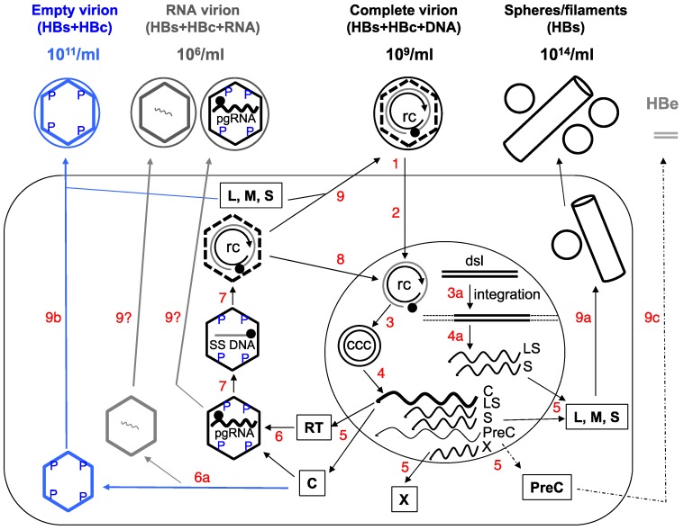
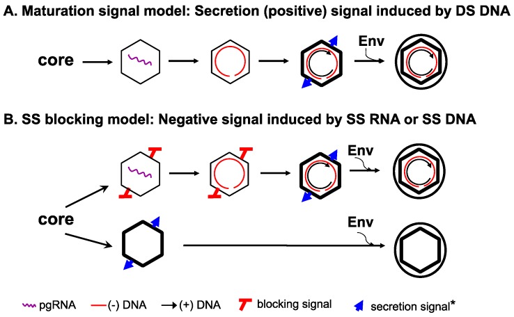
Hepatitis B virus (HBV) is a para-retrovirus or retroid virus that contains a double-stranded DNA genome and replicates this DNA via reverse transcription of a RNA pregenome. Viral reverse transcription takes place within a capsid upon packaging of the RNA and the viral reverse transcriptase. A major characteristic of HBV replication is the selection of capsids containing the double-stranded DNA, but not those containing the RNA or the single-stranded DNA replication intermediate, for envelopment during virion secretion. The complete HBV virion particles thus contain an outer envelope, studded with viral envelope proteins, that encloses the capsid, which, in turn, encapsidates the double-stranded DNA genome. Furthermore, HBV morphogenesis is characterized by the release of subviral particles that are several orders of magnitude more abundant than the complete virions. One class of subviral particles are the classical surface antigen particles (Australian antigen) that contain only the viral envelope proteins, whereas the more recently discovered genome-free (empty) virions contain both the envelope and capsid but no genome. In addition, recent evidence suggests that low levels of RNA-containing particles may be released, after all. We will summarize what is currently known about how the complete and incomplete HBV particles are assembled. We will discuss briefly the functions of the subviral particles, which remain largely unknown. Finally, we will explore the utility of the subviral particles, particularly, the potential of empty virions and putative RNA virions as diagnostic markers and the potential of empty virons as a vaccine candidate.

Keywords: Australian antigen; CCC DNA; HBcAg; HBsAg; diagnosis; empty virion; hepatitis B virus; subviral particles; vaccine; virion.


Conflict of interest statement

The authors declare no conflict of interest.

Figures

Figure 1
Figure 1
Schematic of hepatitis B virus (HBV) replication cycle. 1. Virus binding and entry into the host cell (large rectangle). 2. Intracellular trafficking and delivery of relaxed circular (RC) DNA to the nucleus (large circle). 3. Conversion of RC DNA to CCC DNA, or integration of the double-stranded linear (DSL) DNA into host DNA (3a). 4. and 4a. Transcription to synthesize viral RNAs (wavy lines), including the C mRNA for both the core and RT proteins; LS mRNA for the L envelope protein; S mRNA for the M and S envelope proteins; X mRNA for the X protein; and PreC mRNA for the PreCore protein. The C mRNA is also the pgRNA. 5. Translation to synthesize viral proteins. 6. Assembly of the pgRNA- (and RT-) containing NC, or alternatively, empty capsids (6a). 7. Reverse transcription of pgRNA to synthesize the (−) strand SS DNA and then RC DNA. 8. Nuclear recycling of progeny RC DNA to form more CCC DNA (intracellular CCC DNA amplification). 9. Envelopment of the RC DNA-containing NC and secretion of complete virions, or alternatively, secretion of empty virions (9b) or HBsAg spheres and filaments (9a). Processing of the PreCore protein and secretion of HBeAg are depicted in 9c. The secretion of putative RNA virions is not yet resolved (9?). The different viral particles outside the cell are depicted schematically with their approximate concentrations in the blood of infected persons indicated: the complete, empty, or RNA virions as large circles (outer envelope) with an inner diamond shell (capsid), with or without RC DNA (unclosed, double concentric circle) or RNA (wavy line) inside the capsid respectively; HBsAg spheres and filament as small circles and a cylinder. It is important to point out that the concentrations of all these particles can vary widely between different patients and over time in the same patient. Intracellular capsids are depicted as diamonds, with either viral pgRNA, SS [(−) strand] DNA (straight line), RC DNA, or empty. The letters “P” denote phosphorylated residues on the immature NCs (containing SS DNA or pgRNA) or empty capsid. The dashed lines of the diamond in the RC DNA-containing mature NCs signify the destabilization of the mature NC, which is dephosphorylated. The empty capsids, like mature NCs, are also less stable compared to immature NCs but unlike mature NCs, are phosphorylated. The soluble, dimeric HBeAg is depicted as grey double bars. The thin dashed line and arrow denote the fact that HBeAg is frequently decreased or lost late in infection. Boxed letters denote the viral proteins translated from the mRNAs. The filled circle on RC DNA denotes the RT protein attached to the 5’ end of the (−) strand (outer circle) of RC DNA and the arrow denotes the 3’ end of the (+) strand (inner circle) of RC DNA. ccc, CCC DNA; rc, RC DNA. For simplicity, synthesis of the minor DSL form of the genomic DNA in the mature NC, its secretion in virions, and infection of DSL DNA-containing virions are not depicted here, as are the functions of X. See text for details. Modified from [2].
Figure 2
Figure 2
The single strand blocking hypothesis to explain selective HBV virion formation. The new hypothesis is presented in panel (B), in comparison with the classical maturation signal hypothesis depicted in panel (A). The symbol * denotes that the envelope signal for the mature NC vs. the empty capsid may or may not be the same. See text for details. Modified from [8].

References

    1. Hu J., Seeger C. Hepadnavirus genome replication and persistence. Cold Spring Harb. Perspect. Med. 2015;5:a021386. doi: 10.1101/cshperspect.a021386. - DOI - PMC - PubMed
    1. Hu J. Hepatitis B virus virology and replication. In: Liaw Y.-F., Zoulim F., editors. Hepatitis B Virus in Human Diseases. Humana Press; New York, NY, USA: Dordrecht, The Netherlands: London, UK: 2016. pp. 1–34.
    1. Gerelsaikhan T., Tavis J.E., Bruss V. Hepatitis B virus nucleocapsid envelopment does not occur without genomic DNA synthesis. J. Virol. 1996;70:4269–4274. - PMC - PubMed
    1. Summers J., Mason W.S. Replication of the genome of a hepatitis B--like virus by reverse transcription of an RNA intermediate. Cell. 1982;29:403–415. doi: 10.1016/0092-8674(82)90157-X. - DOI - PubMed
    1. Perlman D., Hu J. Duck hepatitis B virus virion secretion requires a double-stranded DNA genome. J. Virol. 2003;77:2287–2294. doi: 10.1128/JVI.77.3.2287-2294.2003. - DOI - PMC - PubMed

Publication types