. 2017 Mar 23;18(1):16.
doi: 10.1186/s12860-017-0132-1.

Testis-specific ATP synthase peripheral stalk subunits required for tissue-specific mitochondrial morphogenesis in Drosophila

Affiliations

Testis-specific ATP synthase peripheral stalk subunits required for tissue-specific mitochondrial morphogenesis in Drosophila

Eric M Sawyer et al. BMC Cell Biol. .

Abstract

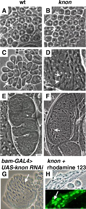
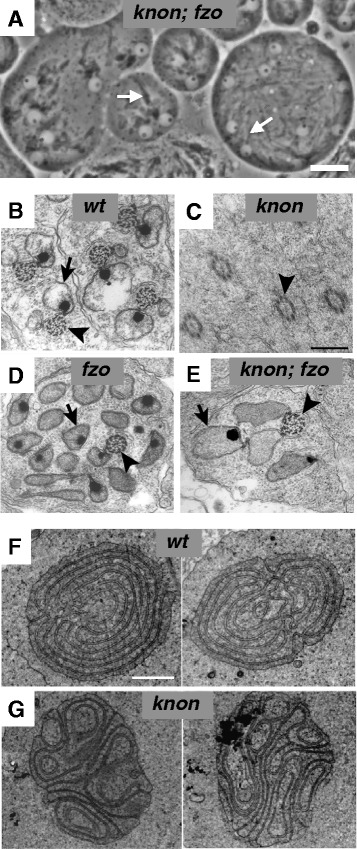
Background: In Drosophila early post-meiotic spermatids, mitochondria undergo dramatic shaping into the Nebenkern, a spherical body with complex internal structure that contains two interwrapped giant mitochondrial derivatives. The purpose of this study was to elucidate genetic and molecular mechanisms underlying the shaping of this structure.

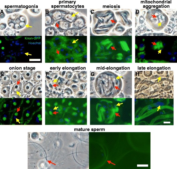
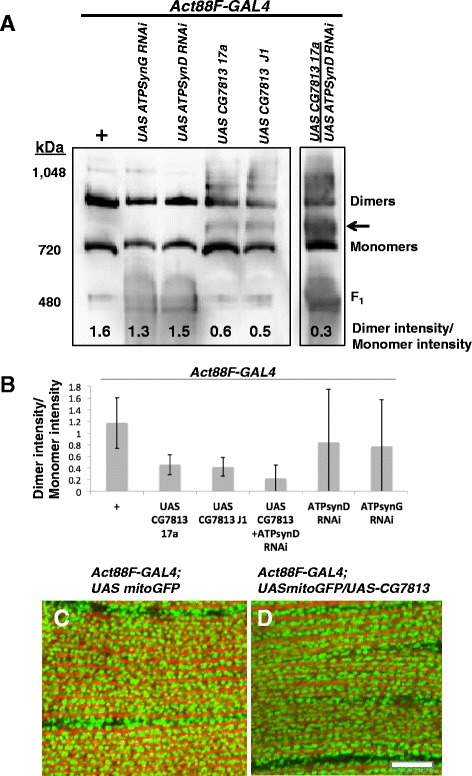
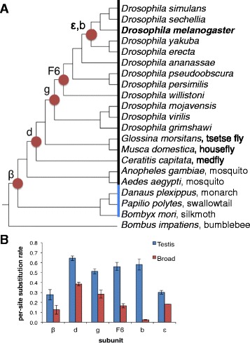
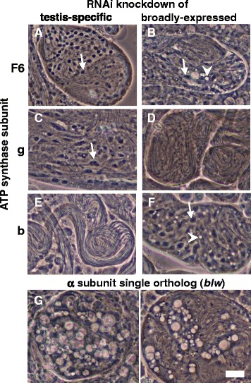
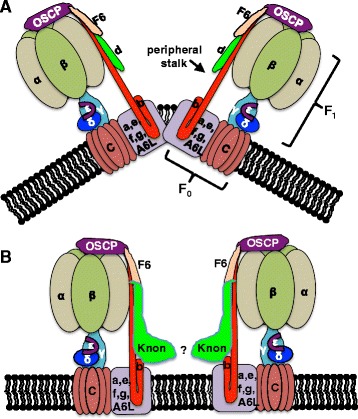
Results: The knotted onions (knon) gene encodes an unconventionally large testis-specific paralog of ATP synthase subunit d and is required for internal structure of the Nebenkern as well as its subsequent disassembly and elongation. Knon localizes to spermatid mitochondria and, when exogenously expressed in flight muscle, alters the ratio of ATP synthase complex dimers to monomers. By RNAi knockdown we uncovered mitochondrial shaping roles for other testis-expressed ATP synthase subunits.

Conclusions: We demonstrate the first known instance of a tissue-specific ATP synthase subunit affecting tissue-specific mitochondrial morphogenesis. Since ATP synthase dimerization is known to affect the degree of inner mitochondrial membrane curvature in other systems, the effect of Knon and other testis-specific paralogs of ATP synthase subunits may be to mediate differential membrane curvature within the Nebenkern.

Keywords: ATP synthase; Cristae; Drosophila melanogaster; Mitochondria; Spermatogenesis.


Figures

Fig. 1
Fig. 1
knon mutant and knockdown spermatids show aberrant Nebenkern elongation but have an intact membrane potential. Phase-contrast micrographs of live squashed testis preparations showing post-meiotic spermatids from wild-type (a, c, and e) and homozygous knon ms(2)1400 males (b, d, f, h) at the early round (onion) stage (a-b), the early elongation stage (c-d), and later in spermatid elongation (e-f). g RNAi knockdown of knon in the testis recapitulates the mutant phenotype, with unelongated Nebenkerne (arrow) in nearly mature elongating spermatid cysts. h Paired phase-contrast and fluorescence micrographs of an elongating spermatid cyst from a knon ms(2)1400 /Df(2R)7E male. Rhodamine 123 uptake by the unelongated mitochondrial derivatives (arrow) in a nearly mature spermatid cyst indicates ongoing respiratory activity. Scale bar 20 μm
Fig. 2
Fig. 2
knon mutants exhibit aberrant Nebenkern structure and defective mitochondrial elongation that can be suppressed by bypassing mitochondrial fusion. a Phase-contrast micrograph of early elongation-stage spermatids from knon ms(2)1400 ; fzo 1 males show some degree of elongation of unfused mitochondria (arrows); compare to Fig. 1c-d. b-e Cross sections of elongating spermatids viewed by transmission electron microscopy showing axonemes (arrowheads) and elongating mitochondrial derivatives (arrows). Wild-type spermatids (b) show two mitochondrial derivatives per axoneme. c knon ms(2)1400 spermatids lack detectable mitochondrial derivatives beside the axoneme. d fzo 1 spermatid has multiple mitochondrial derivatives per axoneme resulting from failure of mitochondrial fusion to form the Nebenkern [3]. e Spermatid from knon ms(2)1400 ; fzo 1 male shows several mitochondrial derivatives elongating beside the axoneme. Transmission electron micrographs of cross sections of Nebenkerne from wild-type (f) and knon ms(2)1400 (g) males. Two examples of each are shown. Internal “onion” structure is disrupted in knon ms(2)1400 . Scale bars 20 μm in a, 0.5 μm in b-e, and 2 μm in f-g. b and d adapted from [3]
Fig. 3
Fig. 3
Knon-GFP rescues the mutant phenotype and localizes to mitochondria in primary spermatocytes and post-meiotic spermatids. Phase-contrast and paired fluorescence images of cells from testes of knon ms(2)1400 /Df(2R)7E; knon-GFP/+ males, stained with Hoechst. Knon-GFP (green) was not detectable in spermatogonia (a) but was associated with phase-dark mitochondria (red arrows) in all later stages (b-i). Yellow arrows indicate nuclei; red arrows indicate mitochondria-associated Knon-GFP. In primary spermatocytes (b), Knon-GFP-labeled mitochondria were small and diffuse in the cytoplasm. In meiotic cells (c), Knon-GFP-marked mitochondria associated with the spindle; immediately after meiosis (d) mitochondria aggregated beside each nucleus and were associated with a strong Knon-GFP signal. The mitochondrial derivatives within the Nebenkern at the onion stage (e, red arrow) and during early- (f) and mid-elongation (g) showed unambiguous Knon-GFP localization. In nearly mature elongating spermatid cysts (h), elongated flagella appear wild type with no clumped mitochondrial derivatives (compare to Fig. 1). Knon-GFP is evenly distributed throughout the elongated spermatids. After sperm individualization, motile sperm (i) retain detectable Knon-GFP (red arrow). The apparent syncytial nature of some cells at earlier stages is an artefact of the preparation—ring canals connecting cells in a cyst are commonly burst open by the pressure from a cover slip. These localization results are identical to those seen when Knon-GFP is expressed in a wild-type background (not shown). Scale bars in a-g, h, and i are 20 μm
Fig. 4
Fig. 4
Knon expression in flight muscle alters ATP synthase dimer:monomer ratio but not gross mitochondrial morphology. a Immunoblot using anti-ATP synthase α subunit antibodies on a BN-PAGE gel of mitochondrial isolates from flight muscle of the indicated genotypes. The Act88F-GAL4 flight muscle driver was used as a control (first lane), as well as to drive exogenous Knon (CG7813) expression from two independent transgene insertions (fourth and fifth lanes), RNAi knockdown constructs of ATP synthase subunits g (second lane) and d (third lane), or simultaneous exogenous CG7813 expression and knockdown of ATP synthase subunit d (sixth lane). Dimer/monomer intensity ratios are indicated at bottom. Expressing Knon (CG7813), either with or without concurrent knockdown of the broadly-expressed ATPsynD, reduced the amount of ATP synthase dimers relative to monomers and gave rise to detectable bands of slightly larger size (arrow) than the typical monomer bands. b Quantification of dimer/monomer band intensity ratios from multiple biological replicates (n = 3 for +, UAS CG7813 17a, and UAS CG7813 J1; n = 4 for UAS CG7813 + ATPsynD RNAi; n = 2 for ATPsynD RNAi and ATPsynG RNAi). Error bars represent 95% confidence intervals. c-d Confocal images of Drosophila flight muscle from Act88F-GAL4; UAS-mitoGFP (c) flies and Act88F-GAL4; UAS-mitoGFP/UAS-CG7813 flies (d). Scale bar 10 μm
Fig. 5
Fig. 5
Phylogeny and evolution of Knon and other ATP synthase subunit paralogs in insects. a The six ATP synthase subunits encoded by two paralogs in D. melanogaster have widely varying phylogenetic origins, determined by comparisons among insect genome sequences (black, Diptera; blue, Lepidoptera) deposited in NCBI. Circles indicate the most recent common ancestor that contained both paralogs. b Testis-enriched subunits have acquired more amino acid substitutions relative to a recent outgroup than broadly expressed subunits. The testis-specific ortholog of each subunit was inferred based on sequence similarity to D. melanogaster orthologs, which were in all cases unambiguous. The average per-site amino acid substitution rate for each subunit type relative to the most recent outgroup of the gene duplication was calculated from a Clustal Omega alignment [76]. Error bars indicate standard deviation among the species analyzed
Fig. 6
Fig. 6
RNAi knockdown of other ATP synthase subunits in the testis. a-f bam-GAL4-driven UAS-RNAi testis knockdown of ATP synthase subunit paralogs that show either testis-enriched expression (left column) or broad expression across multiple tissues (right column). Two of the three testis-enriched paralogs of peripheral stalk subunits—F6 (a) and g (c), but not b (e)—show a similar knockdown phenotype to knon, with failure of mitochondrial elongation (arrows) in spermatid cysts that have nearly fully elongated, along with individualization failure, a common secondary effect of spermatid morphology defects [6]. Knockdown of broadly-expressed paralogs of subunits F6 (b) and b (f) results in aberrant mitochondrial elongation along with vacuolar inclusions (arrowheads), whereas knockdown of broadly-expressed subunit g (d) results in elongating spermatid cysts of wild-type appearance. g Testis knockdown of the sole ATP synthase α subunit ortholog (bellwether), part of the F1 portion of ATP synthase, resulted in large vacuolar inclusions and degradation of elongating spermatid cysts. Two representative cysts are shown. Scale bar 20 μm for a-g. For each genotype, >80 spermatid cysts were visualized from >6 dissected males
Fig. 7
Fig. 7
Model for alteration of ATP synthase complex dimerization by Knon. a Schematic representation of ATP synthase complex dimerization as mediated by various subunits in the peripheral stalk and F0 portion. b Steric interactions of the large Knon C termini might inhibit dimerization or enable dimerization at a shallower angle

Similar articles

Cited by

References

    1. Friedman JR, Nunnari J. Mitochondrial form and function. Nature. 2014;505:335–343. doi: 10.1038/nature12985. - DOI - PMC - PubMed
    1. Mishra P, Chan DC. Mitochondrial dynamics and inheritance during cell division, development and disease. Nat Rev Mol Cell Biol. 2014;15:634–646. doi: 10.1038/nrm3877. - DOI - PMC - PubMed
    1. Hales KG, Fuller MT. Developmentally regulated mitochondrial fusion mediated by a conserved, novel, predicted GTPase. Cell. 1997;90:121–129. doi: 10.1016/S0092-8674(00)80319-0. - DOI - PubMed
    1. Stowers RS, Megeath LJ, Gorska-Andrzejak J, Meinertzhagen IA, Schwarz TL. Axonal transport of mitochondria to synapses depends on milton, a novel Drosophila protein. Neuron. 2002;36:1063–1077. doi: 10.1016/S0896-6273(02)01094-2. - DOI - PubMed
    1. Tates AD. Cytodifferentiation during spermatogenesis in Drosophila melanogaster: an electron microscope study. Leiden: Rijksuniversiteit; 1971.

MeSH terms

Substances

LinkOut - more resources