

The role of pili in Bacillus cereus intraocular infection
- PMID: 28336259
- PMCID: PMC5492386
- DOI: 10.1016/j.exer.2017.03.007
The role of pili in Bacillus cereus intraocular infection
Abstract
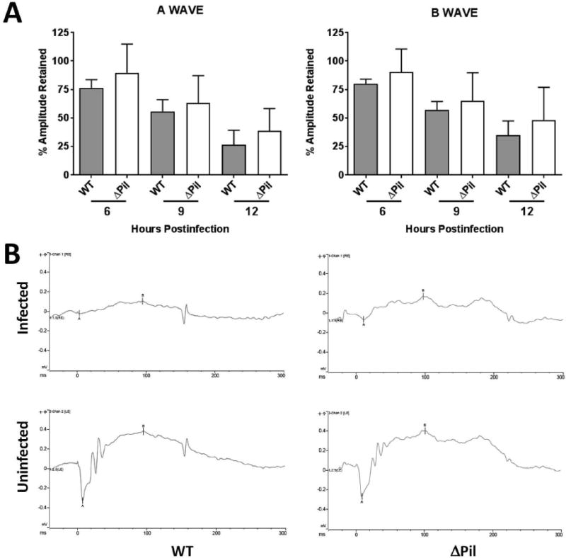
Bacterial endophthalmitis is a potentially blinding intraocular infection. The bacterium Bacillus cereus causes a devastating form of this disease which progresses rapidly, resulting in significant inflammation and loss of vision within a few days. The outer surface of B. cereus incites the intraocular inflammatory response, likely through interactions with innate immune receptors such as TLRs. This study analyzed the role of B. cereus pili, adhesion appendages located on the bacterial surface, in experimental endophthalmitis. To test the hypothesis that the presence of pili contributed to intraocular inflammation and virulence, we analyzed the progress of experimental endophthalmitis in mouse eyes infected with wild type B. cereus (ATCC 14579) or its isogenic pilus-deficient mutant (ΔbcpA-srtD-bcpB or ΔPil). One hundred CFU were injected into the mid-vitreous of one eye of each mouse. Infections were analyzed by quantifying intraocular bacilli and retinal function loss, and by histology from 0 to 12 h postinfection. In vitro growth and hemolytic phenotypes of the infecting strains were also compared. There was no difference in hemolytic activity (1:8 titer), motility, or in vitro growth (p > 0.05, every 2 h, 0-18 h) between wild type B. cereus and the ΔPil mutant. However, infected eyes contained greater numbers of wild type B. cereus than ΔPil during the infection course (p ≤ 0.05, 3-12 h). Eyes infected with wild type B. cereus experienced greater losses in retinal function than eyes infected with the ΔPil mutant, but the differences were not always significant. Eyes infected with ΔPil or wild type B. cereus achieved similar degrees of severe inflammation. The results indicated that the intraocular growth of pilus-deficient B. cereus may have been better controlled, leading to a trend of greater retinal function in eyes infected with the pilus-deficient strain. Although this difference was not enough to significantly alter the severity of the inflammatory response, these results suggest a potential role for pili in protecting B. cereus from clearance during the early stages of endophthalmitis, which is a newly described virulence mechanism for this organism and this infection.
Keywords: Bacillus; Endophthalmitis; Eye; Infection; Inflammation; Pili; Virulence.
Copyright © 2017 Elsevier Ltd. All rights reserved.
Figures


Similar articles
-
Role of Toll-like receptor (TLR) 2 in experimental Bacillus cereus endophthalmitis.PLoS One. 2011;6(12):e28619. doi: 10.1371/journal.pone.0028619. Epub 2011 Dec 6. PLoS One. 2011. PMID: 22163046 Free PMC article.
-
Role of TLR5 and flagella in bacillus intraocular infection.PLoS One. 2014 Jun 24;9(6):e100543. doi: 10.1371/journal.pone.0100543. eCollection 2014. PLoS One. 2014. PMID: 24959742 Free PMC article.
-
Role of swarming migration in the pathogenesis of bacillus endophthalmitis.Invest Ophthalmol Vis Sci. 2006 Oct;47(10):4461-7. doi: 10.1167/iovs.06-0301. Invest Ophthalmol Vis Sci. 2006. PMID: 17003440
-
The cereus matter of Bacillus endophthalmitis.Exp Eye Res. 2020 Apr;193:107959. doi: 10.1016/j.exer.2020.107959. Epub 2020 Feb 4. Exp Eye Res. 2020. PMID: 32032628 Free PMC article. Review.
-
Posttraumatic endophthalmitis: the emerging role of Bacillus cereus infection.Rev Infect Dis. 1987 Jan-Feb;9(1):110-23. doi: 10.1093/clinids/9.1.110. Rev Infect Dis. 1987. PMID: 3103191 Review.
Cited by
-
Roles of CCL2 and CCL3 in intraocular inflammation during Bacillus endophthalmitis.Exp Eye Res. 2022 Nov;224:109213. doi: 10.1016/j.exer.2022.109213. Epub 2022 Sep 2. Exp Eye Res. 2022. PMID: 36063964 Free PMC article.
-
Bacillus S-Layer-Mediated Innate Interactions During Endophthalmitis.Front Immunol. 2020 Feb 12;11:215. doi: 10.3389/fimmu.2020.00215. eCollection 2020. Front Immunol. 2020. PMID: 32117322 Free PMC article.
-
The Bacillus virulome in endophthalmitis.Microbiology (Reading). 2021 May;167(5):001057. doi: 10.1099/mic.0.001057. Microbiology (Reading). 2021. PMID: 34032564 Free PMC article.
-
Immune Inhibitor A Metalloproteases Contribute to Virulence in Bacillus Endophthalmitis.Infect Immun. 2021 Sep 16;89(10):e0020121. doi: 10.1128/IAI.00201-21. Epub 2021 Jun 7. Infect Immun. 2021. PMID: 34097460 Free PMC article.
-
C-X-C Chemokines Influence Intraocular Inflammation During Bacillus Endophthalmitis.Invest Ophthalmol Vis Sci. 2021 Nov 1;62(14):14. doi: 10.1167/iovs.62.14.14. Invest Ophthalmol Vis Sci. 2021. PMID: 34784411 Free PMC article.
References
-
- Abdul-Careem MF, Firoz Mian M, Gillgrass AE, Chenoweth MJ, Barra NG, Chan T, Al-Garawi AA, Chew MV, Yue G, van Roojen N, Xing Z, Ashkar AA. FimH, a TLR4 ligand, induces innate antiviral responses in the lung leading to protection against lethal influenza infection in mice. Antiviral Res. 2011;92:346–355. - PubMed
-
- Axner O, Andersson M, Björnham O, Castelain M, Klinth J, Koutris E, Schedin S. Assessing bacterial adhesion on an individual adhesin and single pili level using optical tweezers. Adv Exp Med Biol. 2011;715:301–313. - PubMed
-
- Bagnoli F, Moschioni M, Donati C, Dimitrovska V, Ferlenghi I, Facciotti C, Muzzi A, Giusti F, Emolo C, Sinisi A, Hilleringmann M, Pansegrau W, Censini S, Rappuoli R, Covacci A, Masignani V, Barocchi MA. A second pilus type in Streptococcus pneumoniae is prevalent in emerging serotypes and mediates adhesion to host cells. J Bacteriol. 2008;190:5480–5492. - PMC - PubMed
Publication types
MeSH terms
Grants and funding
LinkOut - more resources
Full Text Sources
Other Literature Sources
Molecular Biology Databases
