Review
. 2017 Sep:60:201-218.
doi: 10.1016/j.preteyeres.2017.03.002. Epub 2017 Mar 20.

The impact of oxidative stress and inflammation on RPE degeneration in non-neovascular AMD

Affiliations
Review

The impact of oxidative stress and inflammation on RPE degeneration in non-neovascular AMD

Sayantan Datta et al. Prog Retin Eye Res. 2017 Sep.

Abstract

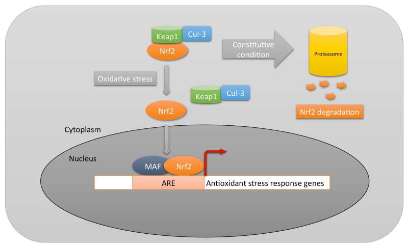
The retinal pigment epithelium (RPE) is a highly specialized, unique epithelial cell that interacts with photoreceptors on its apical side and with Bruch's membrane and the choriocapillaris on its basal side. Due to vital functions that keep photoreceptors healthy, the RPE is essential for maintaining vision. With aging and the accumulated effects of environmental stresses, the RPE can become dysfunctional and die. This degeneration plays a central role in age-related macular degeneration (AMD) pathobiology, the leading cause of blindness among the elderly in western societies. Oxidative stress and inflammation have both physiological and potentially pathological roles in RPE degeneration. Given the central role of the RPE, this review will focus on the impact of oxidative stress and inflammation on the RPE with AMD pathobiology. Physiological sources of oxidative stress as well as unique sources from photo-oxidative stress, the phagocytosis of photoreceptor outer segments, and modifiable factors such as cigarette smoking and high fat diet ingestion that can convert oxidative stress into a pathological role, and the negative impact of impairing the cytoprotective roles of mitochondrial dynamics and the Nrf2 signaling system on RPE health in AMD will be discussed. Likewise, the response by the innate immune system to an inciting trigger, and the potential role of local RPE production of inflammation, as well as a potential role for damage by inflammation with chronicity if the inciting trigger is not neutralized, will be debated.

Keywords: Age-related macular degeneration; Complement; Inflammation; Mitochondrial dynamics; Nrf2; Oxidative stress; Retinal pigment epithelium.


Conflict of interest statement

Conflict of Interest: JTH and MC have received funding from Bayer Pharmaceuticals, Inc.

Figures

Figure 1
Figure 1
Diagram of Nrf2 signaling. In the absence of oxidative stress (ROS), Nrf2 is degraded through the proteasome by a Keap1/Cul3 mechanism. With oxidative stress, the ROS induce a conformational change in cytoplasmic Keap1, which releases Nrf2 for transit to the nucleus where it interacts with Maf proteins and binds to antioxidant response elements (ARE), inducing a comprehensive antioxidant response.
Figure 2
Figure 2
Nrf2 immunolabeling in a 60-year-old Caucasian female with early AMD. A) Macular RPE with normal, cuboidal morphology have prominent cytoplasmic Nrf2 labeling (blue). B) IgG control. C) Normal macular RPE with both cytoplasmic and nuclear Nrf2 labeling (arrowheads). D) Dysmorphic macular RPE overlying drusen have lighter cytoplasmic labeling than cuboidal RPE from the same section. Nuclei (arrowheads) do not stain for Nrf2. E) Peripheral RPE with minimal Nrf2 labeling in the cytoplasm and no nuclear labeling (arrowheads). F–H) Same images as C–E, respectively, after Nuance software has converted melanin to maroon to improve visualization of Nrf2 labeling in the RPE. The arrow points to the inset of an enlarged image showing nuclear Nrf2 staining in a normal macular RPE cell (F), lack of nuclear Nrf2 staining in a dysmorphic macular RPE cell overlying a druse (*) (G), and lack of Nrf2 staining in a peripheral RPE cell (H). CH, choroid, RPE, retinal pigment epithelium, Black arrows point to Bruch’s membrane. Bar=25μm. Reprinted with permission from Free Radical Biology Medicine.
Figure 3
Figure 3
Distribution of CD46 in the RPE of early AMD eyes. A. Peripheral RPE/choroid from a 94 yr. old Caucasian female with early AMD. CD46 labeling appears as a prominent line at the basal (arrowhead) and lateral (arrows) surfaces of morphologically normal RPE cells. C. Dysmorphic macular RPE cells, from the same patient, adjacent to small druse with reduced (thinner line) or undetectable CD46 labeling at their lateral surface (arrows). The basal line of CD46 labeling (arrowhead) is preserved, but thinned over the small druse. E. 96 year old Caucasian male with early AMD and a large druse (>500 μm in length). RPE cells overlying the large druse are dysmorphic and the uniform line of lateral (arrows) and basal (arrowhead) CD46 labeling is lost. CD46 labeling is diffusely distributed throughout the druse (*). G. IgG control. B, D, F, H. Same images after RPE melanin pigment is subtracted by Nuance software. Ch, choroid; Dr, druse; RPE, retinal pigmented epithelium. Bar=50μm. Reprinted with permission from Journal of Pathology.
Figure 4
Figure 4
Immunofluorescence confocal microscopy of a WT mouse given IVit vehicle (n=4) shows linear FH (red) staining (i) and PTX3 (green) staining (ii) in inner BrM (white arrowheads). (iii) Merged image of (i) and (ii) shows FH and PTX3 (yellow) staining overlap. WT mouse given 5 μg 4-HNE IVit (n=4) has diffuse staining in the RPE for FH (red) (iv) and PTX3 (green) (v). (vi) Merged image of (iv) and (v) shows FH and PTX3 (yellow) staining overlap. IgG isotype control for FH (vii) and PTX3 (viii). Reprinted with permission from Journal of Pathology.
Figure 5
Figure 5
Diagram of the alternative complement pathway and the inflammasome. Activation of the alternative complement pathway is regulated by factor H (CFH), CD46, CD55. PTX3, by binding CFH, controls the abundance of CFH. Besides its regulation of complement, CFH binds to malondialdehyde (MDA) to control MDA-mediated pro-inflammatory cytokine production, and heparan sulfates (HS) to compete with very low density lipoproteins (VLDL) that can become deposited in Bruch’s membrane prior to basal deposit or drusen formation. Complement activation can lead to anaphylatoxins generation of C3a and C5a, which can induce pro-inflammatory cytokine production, and C5b-9 complex formation, which is regulated by CD59. C3a, C5a, and C5b-9 complexes are implicated in activating the inflammasome, composed of NLRP3, ASP, and Pro-caspase1, which upon activation, is cleaved to convert pro-IL-1β, pro-IL-18, and pro-IL-33 into their active forms.

References

    1. Lutein + zeaxanthin and omega-3 fatty acids for age-related macular degeneration: the Age-Related Eye Disease Study 2 (AREDS2) randomized clinical trial. JAMA. 309:2005–2015. - PubMed
    1. Abdelsalam A, Del Priore L, Zarbin MA. Drusen in age-related macular degeneration: pathogenesis, natural course, and laser photocoagulation-induced regression. Surv Ophthalmol. 1999;44:1–29. - PubMed
    1. Ach T, Huisingh C, McGwin G, Jr, Messinger JD, Zhang T, Bentley MJ, Gutierrez DB, Ablonczy Z, Smith RT, Sloan KR, Curcio CA. Quantitative autofluorescence and cell density maps of the human retinal pigment epithelium. Invest Ophthalmol Vis Sci. 2014;55:4832–4841. - PMC - PubMed
    1. Ach T, Tolstik E, Messinger JD, Zarubina AV, Heintzmann R, Curcio CA. Lipofuscin redistribution and loss accompanied by cytoskeletal stress in retinal pigment epithelium of eyes with age-related macular degeneration. Invest Ophthalmol Vis Sci. 2015;56:3242–3252. - PMC - PubMed
    1. Adams J, Kelso R, Cooley L. The kelch repeat superfamily of proteins: propellers of cell function. Trends in cell biology. 2000;10:17–24. - PubMed

Publication types

MeSH terms