. 2017 Mar 24;355(6331):1312-1317.
doi: 10.1126/science.aad8242.

A conserved NAD+ binding pocket that regulates protein-protein interactions during aging

Affiliations

A conserved NAD+ binding pocket that regulates protein-protein interactions during aging

Jun Li et al. Science. .

Abstract

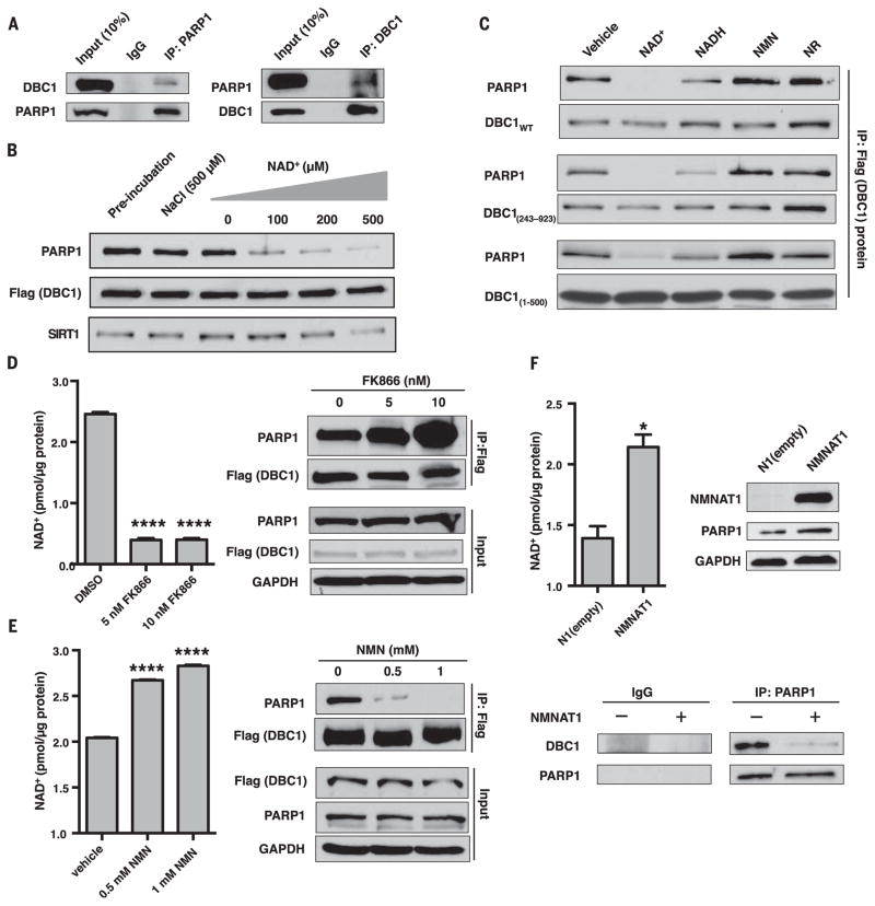
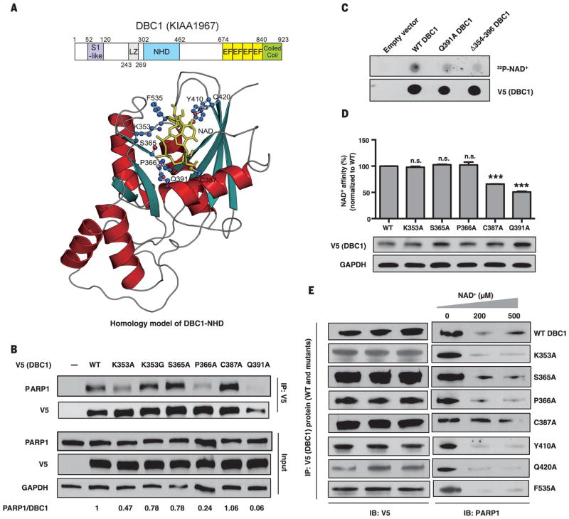
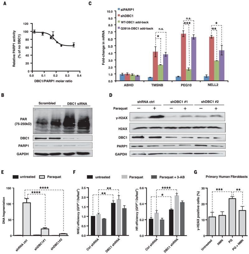
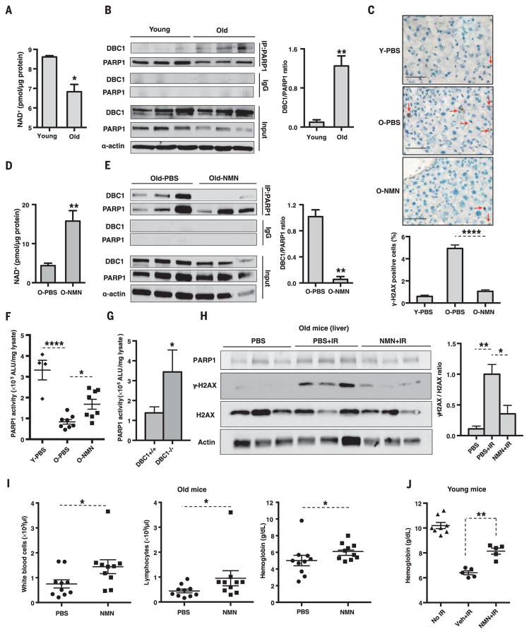
DNA repair is essential for life, yet its efficiency declines with age for reasons that are unclear. Numerous proteins possess Nudix homology domains (NHDs) that have no known function. We show that NHDs are NAD+ (oxidized form of nicotinamide adenine dinucleotide) binding domains that regulate protein-protein interactions. The binding of NAD+ to the NHD domain of DBC1 (deleted in breast cancer 1) prevents it from inhibiting PARP1 [poly(adenosine diphosphate-ribose) polymerase], a critical DNA repair protein. As mice age and NAD+ concentrations decline, DBC1 is increasingly bound to PARP1, causing DNA damage to accumulate, a process rapidly reversed by restoring the abundance of NAD+ Thus, NAD+ directly regulates protein-protein interactions, the modulation of which may protect against cancer, radiation, and aging.


Figures

Fig. 1
Fig. 1. Regulation of the PARP1-DBC1 interaction by NAD+
(A) Endogenous DBC1 and PARP1 interact. IgG, immunoglobulin G; IP, immunoprecipitation. (B) NAD+ dissociates the PARP1-DBC1 interaction. (C) Effects of NAD+ and structurally related molecules on the PARP1-DBC1 interaction. Flag-DBC1 was incubated with molecules (200 μM) for 1 hour and then probed for PARP1. NMN, nicotinamide mononucleotide; NR, nicotinamide riboside; WT, wild type. (D to F) The PARP1-DBC1 interaction after treatment with (D) FK866 or (E) NMN for 24 hours or (F) in cells overexpressing NMNAT1, an NAD+ salvage pathway gene to raise NAD+. DMSO, dimethyl sulfoxide; GAPDH, glyceraldehyde-3-phosphate dehydrogenase. Error bars indicate mean ± SEM. (D and E) One-way analysis of variance (ANOVA) and Sidak’s post-hoc correction. (F) Unpaired two-tailed t test. *P < 0.05, ****P < 0.0001.
Fig. 2
Fig. 2. Binding of the Nudix homology domain (NHD) of DBC1 to NAD+ and PARP1
(A) Domains and crystallographic-based homology model of the NHD docked with NAD+. S1-like, ribosomal protein S1 OB-fold domain-like; EF, EF-hand; LZ, leucine zipper. Residues predicted to be in the vicinity of bound NAD+ are highlighted. (B) Interaction of V5/His-tagged DBC1 mutants and PARP1. See fig. S8 for additional mutants. (C and D) Direct binding of NAD+ to the DBC1-NHD, assessed using a radiolabeled NAD+ binding assay (C) or a biotin-NAD+ binding assay (D). Error bars indicate mean ± SEM; one-way ANOVA and Sidak’s post-hoc correction. ***P < 0.001; n.s., not significant. (E) Effect of NAD+ on binding of DBC1-NHD mutants to PARP1. Single-letter abbreviations for the amino acid residues are as follows: A, Ala; C, Cys; F, Phe; G, Gly; K, Lys; P, Pro; Q, Gln; S, Ser; and Y, Tyr.
Fig. 3
Fig. 3. DBC1 inhibits PARP1 activity and DNA repair
(A) Inhibition of PARP1 activity by DBC1 purified from HEK 293T cells. (B) PAR [poly(ADP-ribose)] abundance in HEK 293T cells lacking DBC1. siRNA, small interfering RNA. (C) Opposing effects of reintroducing the wild type or DBC1Q391A into MCF-7 cells on mRNA of PARP1-regulated genes. TMSNB, Thymosin beta; PEG10, paternally expressed gene 10; NELL2, neural EGFL-like 2. ABHD2 was a negative control. DBC1 and PARP1 abundance are shown in fig. S10E. sh, short hairpin. (D) γH2AX abundance in DBC1 knockdown cells after paraquat treatment (1 mM, 24 hours). (E) DNA fragmentation after paraquat treatment (0.5 mM, 24 hours) in DBC1 knockdown cells, assessed by a comet assay (>50 cells per group). See fig. S11A. (F) DNA break repair [nonhomologous end joining (NHEJ) and homologous recombination (HR)] in DBC1 knockdown cells treated with paraquat (1 mM) or 3-AB (5 mM). n = 3 biological replicates. (G) Protection of human primary fibroblasts from paraquat (PQ)–induced DNA damage (300 μM paraquat) by NMN (500 μM). 100 ± 20 cells per condition, n = 4 biological replicates (two cell lines, twice for each), 24-hour treatment. See fig. S12. Error bars indicate SEM; one-way ANOVA [(C) and (G)] and two-way ANOVA [(E) and (F)], Sidak’s post-hoc correction. *P < 0.05, **P < 0.01, ***P < 0.001, ****P < 0.0001; n.s., not significant.
Fig. 4
Fig. 4. Increases in the PARP1-DBC1 complex and DNA damage with age are reversed by NMN, a precursor to NAD+
(A and B) NAD+ concentrations and PARP1-DBC1 interactions in livers of young and old mice (6 versus 22 months, n = 3 per group). (C) γH2AX-positive cells (red arrow) in the livers of young (6 months, Y) and old (30 months, O) mice (n = 3 per group) treated for 7 days with vehicle [phosphate-buffered saline (PBS)] or NMN (500 mg/kg per day intraperitoneally, n = 3 per group). Scale bars, 50 μm. (D and E) NAD+ concentrations and PARP1-DBC1 interactions in the livers of old mice (22 months) treated as in (C). (F) PARP1 activity in young (6 months, n = 4) and old (26 ± 4 months, n = 8 per group) mice. (G) PARP1 activity in 18- to 20-month-old DBC1 knockout mice livers (n = 3 or 4). (H) γH2AX abundance in the livers of 26-month-olds after irradiation (IR) [7.5 grays (Gy), 137CsCl], treated as in (C) (n = 3 per group). (I) Blood counts of 23-month-olds on the 7th or 8th day after irradiation (n = 10 per group). See fig. S18, A and B. (J) Blood metrics of 4-month-olds given a single oral dose of NMN (2000 mg/kg) 1 hour after irradiation (8 Gy, 137CsCl), followed by another 7 days of treatment (2000 mg/kg per day, n = 5 to 8 per group). Veh, vehicle. See fig. S18C. Errors bars indicate SEM; unpaired two-tailed t test [(A), (B), (D), (E), and (G)], one-way ANOVA [(C), (F), and (H)], Sidak’s post-hoc correction, Mann-Whitney U-test [(I) and (J)]. *P < 0.05, **P < 0.01, ****P < 0.0001.

References

    1. Houtkooper RH, Cantó C, Wanders RJ, Auwerx J. Endocr Rev. 2010;31:194–223. - PMC - PubMed
    1. Bonkowski MS, Sinclair DA. Nat Rev Mol Cell Biol. 2016;17:679–690. - PMC - PubMed
    1. Imai S, Guarente L. Trends Cell Biol. 2014;24:464–471. - PMC - PubMed
    1. Mouchiroud L, et al. Cell. 2013;154:430–441. - PMC - PubMed
    1. Yoshino J, Mills KF, Yoon MJ, Imai S. Cell Metab. 2011;14:528–536. - PMC - PubMed

Publication types

MeSH terms