. 2017 Jun;174(2):1167-1176.
doi: 10.1104/pp.16.00074. Epub 2017 Mar 23.

GmILPA1, Encoding an APC8-like Protein, Controls Leaf Petiole Angle in Soybean

Affiliations

GmILPA1, Encoding an APC8-like Protein, Controls Leaf Petiole Angle in Soybean

Jinshan Gao et al. Plant Physiol. 2017 Jun.

Abstract

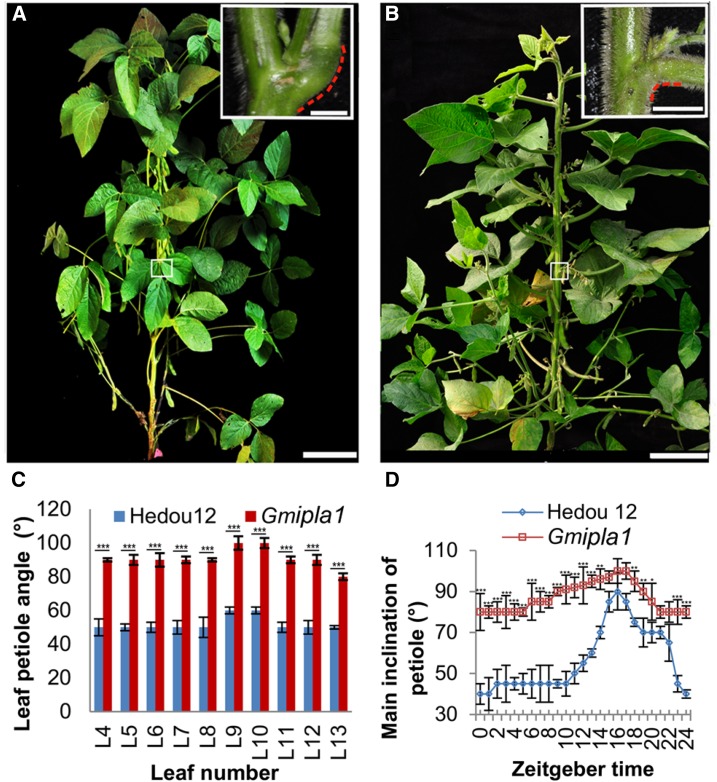
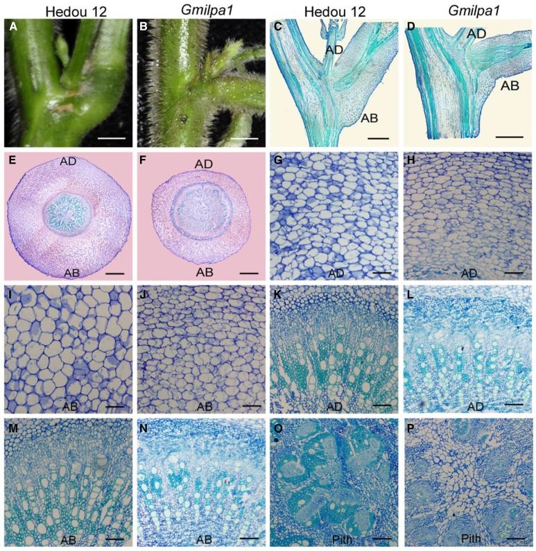
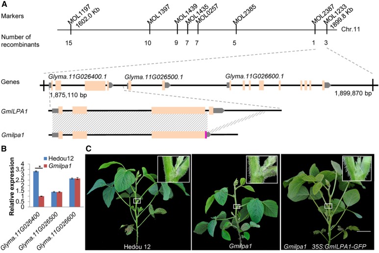
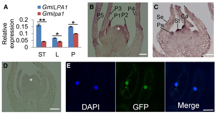
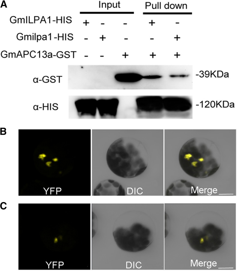
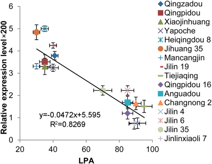
Leaf petiole angle (LPA) is an important plant architectural trait that affects canopy coverage, photosynthetic efficiency, and ultimately productivity in many legume crops. However, the genetic basis underlying this trait remains unclear. Here, we report the identification, isolation, and functional characterization of Glycine max Increased Leaf Petiole Angle1 (GmILPA1), a gene encoding an APC8-like protein, which is a subunit of the anaphase-promoting complex/cyclosome in soybean (Glycine max). A gamma ray-induced deletion of a fragment involving the fourth exon of GmILPA1 and its flanking sequences led to extension of the third exon and formation of, to our knowledge, a novel 3'UTR from intronic and intergenic sequences. Such changes are responsible for enlarged LPAs that are associated with reduced motor cell proliferation in the Gmilpa1 mutant. GmILPA1 is mainly expressed in the basal cells of leaf primordia and appears to function by promoting cell growth and division of the pulvinus that is critical for its establishment. GmILPA1 directly interacts with GmAPC13a as part of the putative anaphase-promoting complex. GmILPA1 exhibits variable expression levels among varieties with different degrees of LPAs, and expression levels are correlated with the degrees of the LPAs. Together, these observations revealed a genetic mechanism modulating the plant petiole angle that could pave the way for modifying soybean plant architecture with optimized petiole angles for enhanced yield potential.


Figures

Figure 1.
Figure 1.
Phenotype of Hedou 12 and the Gmilpa1 mutant. A, Hedou 12; B, the Gmilpa1 mutant at the R5 stage (reproductive stage with approximately 3.2-mm-long seeds in the pod at one of the four uppermost nodes on the main stem). Scale bars, 15 cm. Scale bars in top-right boxes, 1 cm. C, LPAs of the fourth to 13th leaf of Hedou 12 and the Gmilpa1 plants at the R5 stage. LPAs are the means ± ses of the means from 12 different plants. ***P < 0.001 (t test). D, Nyctinastic movement of pulvini in Hedou 12 and the Gmilpa1 mutant. Angles are the means ± ses of the means from 12 different plants. ***P < 0.001, **P < 0.01, *P < 0.05 (t test).
Figure 2.
Figure 2.
Structural comparison of pulvini of 6-week-old Hedou 12 and the Gmilpa1 mutant. A and B, Pulvini of Hedou 12 (A) and the Gmilpa1 mutant (B), respectively. Scale bars, 1 cm. C and D, Longitudinal section of pulvini in Hedou 12 (C) and the Gmilpa1 mutant (D), respectively. Scale bars, 1 cm. E and F, Transection of pulvini in Hedou 12 (E) and the Gmilpa1 mutant (F), respectively. Scale bars, 400 μm. G to P, Partial magnification of transection of pulvini in Hedou 12 (G, K, and O) and the Gmilpa1 mutant (H, L, and P). Scale bars, 50 μm. G and H, The motor cells of Hedou 12 (G) and the Gmilpa1 mutant (H) on the adaxial side. I and J, The motor cells of Hedou 12 (I) and the Gmilpa1 mutant (J) on the abaxial side. K and L, The vascular cylinders of Hedou 12 (K) and the Gmilpa1 mutant (L) on the adaxial side. M and N, The vascular cylinders of Hedou 12 (M) and the Gmilpa1 mutant (N) on the abaxial side. O and P, The piths of Hedou 12 (O) and the Gmilpa1 mutant (P).
Figure 3.
Figure 3.
Map-based cloning of the GmILPA1 locus. A, Physical locations of markers defining the GmILPA1 region, the deletion region in Glyma.11G026400.1, and a new transcript identified in the Gmilpa1 mutant. Shading indicates identical sequences. The chromosomal positions of MOL1197 and MOL1233 are 1602.0 kb and 1890.8 kb, respectively (Glycine max Wm82.a2.v1). B, Expression levels of Glyma.11G026400.1, Glyma.11G026500.1, and Glyma.11G026600.1 in the fifth pulvini at the V5 stage of Hedou 12 and the Gmilpa1 mutant. Expression levels are presented as the means ± ses of the means from four biological replicates. *P < 0.05 (t test). C, Complementation of the Gmilpa1 mutant. Phenotypes of Hedou 12, the Gmilpa1 mutant, and T2 plants with the GmILPA1 transgene. Scale bar, 10 cm. Boxes in the top-right corner illustrate part of plants magnified 10×.
Figure 4.
Figure 4.
Expression and subcellular localization analysis of GmILPA1. A, Expression of GmILPA1 and Gmilpa1 in apical stem tips, leaves, and pulvini in Hedou 12 and the Gmilpa1 mutant. Expression levels are presented as the means ± ses of the means from four biological replicates. **P < 0.01 (t test). B to D, RNA in situ hybridization of GmILPA1 performed using RNA probes in anti-sense (B and C) and sense (D) directions. B and D, The shoot tips were embedded in paraffin at the VE developmental stage (12 d after planting). Asterisks denote the shoot apical meristem. Scale bars, 50 μm. C, The shoot tips were embedded in paraffin at the R1 stage (one open flower at any node). Scale bars, 50 μm. E, Subcellular localization of the GmILPA1-GFP fusion protein in onion epidermal cells under the control of the 35S promoter observed under dark field for green fluorescence (middle). The nuclei were counterstained with 4,6-diamidino-2-phenylindole. Scale bar, 50 μm. Ca, carpel primordium; DAPI, 4,6-diamidino-2-phenylindole; L, leaves; LP, leaf primordium; P, pulvini; Pe, petal primordium; Se, sepal primordium; St, stamen; ST, stem tips.
Figure 5.
Figure 5.
Interaction between GmILPA1/Gmilpa1 and GmAPC13a. A, Interaction between GmILPA1/Gmilpa1 and GmAPC13a detected by HIS-antibody and GST-antibody in the pull-down assays. The “+” and “-” indicate reactions with or without tagged proteins, respectively. B and C, Interaction among GmILPA1 (B), Gmilpa1 (C), and GmAPC13a revealed by BiFC. Scale bars, 10 μm. α-GST, GST-antibody; α-HIS, HIS-antibody.
Figure 6.
Figure 6.
Association analysis of GmILPA1 expression and LPAs in 16 soybean cultivars. LPAs are the means ± ses from six different plants. Expression levels of GmILPA1, analyzed using young leaves at the V5 stage, are the means ± ses from six different plants. Horizontal and vertical bars are the ses of LPA and GmILPA1 expression level × 200, respectively. A significant correlation between the expression levels of GmILPA1 (y) and LPA (x) was found; the trend line was y = −0.0472x + 5.595 (R2 = 0.8269).

Similar articles

Cited by

References

    1. Capron A, Serralbo O, Fülöp K, Frugier F, Parmentier Y, Dong A, Lecureuil A, Guerche P, Kondorosi E, Scheres B, Genschik P (2003) The Arabidopsis anaphase-promoting complex or cyclosome: molecular and genetic characterization of the APC2 subunit. Plant Cell 15: 2370–2382 - PMC - PubMed
    1. Chen J, Moreau C, Liu Y, Kawaguchi M, Hofer J, Ellis N, Chen R (2012) Conserved genetic determinant of motor organ identity in Medicago truncatula and related legumes. Proc Natl Acad Sci USA 109: 11723–11728 - PMC - PubMed
    1. Cheng W, Gao JS, Feng XX, Shao Q, Yang SX, Feng XZ (2016) Characterization of dwarf mutants and molecular mapping of a dwarf locus in soybean. J Integr Agric 15: 60345–60347
    1. Clough SJ, Bent AF (1998) Floral dip: a simplified method for Agrobacterium-mediated transformation of Arabidopsis thaliana. Plant J 16: 735–743 - PubMed
    1. Cortizo M, Laufs P (2012) Genetic basis of the “sleeping leaves” revealed. Proc Natl Acad Sci USA 109: 11474–11475 - PMC - PubMed

LinkOut - more resources