Light Emission Intensities of Luminescent Y₂O₃:Eu and Gd₂O₃:Eu Particles of Various Sizes
- PMID: 28336860
- PMCID: PMC5333011
- DOI: 10.3390/nano7020026
Light Emission Intensities of Luminescent Y₂O₃:Eu and Gd₂O₃:Eu Particles of Various Sizes
Abstract
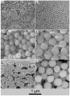
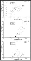
There is great technological interest in elucidating the effect of particle size on the luminescence efficiency of doped rare earth oxides. This study demonstrates unambiguously that there is a size effect and that it is not dependent on the calcination temperature. The Y₂O₃:Eu and Gd₂O₃:Eu particles used in this study were synthesized using wet chemistry to produce particles ranging in size between 7 nm and 326 nm and a commercially available phosphor. These particles were characterized using three excitation methods: UV light at 250 nm wavelength, electron beam at 10 kV, and X-rays generated at 100 kV. Regardless of the excitation source, it was found that with increasing particle diameter there is an increase in emitted light. Furthermore, dense particles emit more light than porous particles. These results can be explained by considering the larger surface area to volume ratio of the smallest particles and increased internal surface area of the pores found in the large particles. For the small particles, the additional surface area hosts adsorbates that lead to non-radiative recombination, and in the porous particles, the pore walls can quench fluorescence. This trend is valid across calcination temperatures and is evident when comparing particles from the same calcination temperature.
Keywords: Gd2O3:Eu; Y2O3:Eu; cathodoluminescence; fluorescence; luminescence; particle size effect; scintillation.
Conflict of interest statement
The authors declare no conflict of interest. The founding sponsors had no role in the design of the study; in the collection, analyses, or interpretation of data; in the writing of the manuscript, and in the decision to publish the results.
Figures


Similar articles
-
Influence of calcination temperature on the luminescent properties of Eu³⁺ doped CaAl₄O₇ phosphor prepared by Pechini method.Spectrochim Acta A Mol Biomol Spectrosc. 2015 Jan 5;134:283-7. doi: 10.1016/j.saa.2014.06.023. Epub 2014 Jun 26. Spectrochim Acta A Mol Biomol Spectrosc. 2015. PMID: 25022500
-
Rare-earth doped gadolinia based phosphors for potential multicolor and white light emitting deep UV LEDs.Nanotechnology. 2009 Mar 25;20(12):125707. doi: 10.1088/0957-4484/20/12/125707. Epub 2009 Mar 4. Nanotechnology. 2009. PMID: 19420484
-
Bio-inspired synthesis of Y2O3: Eu(3+) red nanophosphor for eco-friendly photocatalysis.Spectrochim Acta A Mol Biomol Spectrosc. 2015 Apr 15;141:149-60. doi: 10.1016/j.saa.2015.01.055. Epub 2015 Jan 31. Spectrochim Acta A Mol Biomol Spectrosc. 2015. PMID: 25668696
-
Red emission phosphor for real-time skin dosimeter for fluoroscopy and interventional radiology.Med Phys. 2014 Oct;41(10):101913. doi: 10.1118/1.4893534. Med Phys. 2014. PMID: 25281965
-
Laser excited long lasting luminescence in CaAl₂O₄: Eu³⁺/Eu²⁺+Nd³⁺ phosphor.Spectrochim Acta A Mol Biomol Spectrosc. 2013 Feb;102:212-8. doi: 10.1016/j.saa.2012.09.054. Epub 2012 Oct 13. Spectrochim Acta A Mol Biomol Spectrosc. 2013. PMID: 23220659
Cited by
-
ZnWO4 Nanoparticle Scintillators for High Resolution X-ray Imaging.Nanomaterials (Basel). 2020 Aug 31;10(9):1721. doi: 10.3390/nano10091721. Nanomaterials (Basel). 2020. PMID: 32878007 Free PMC article.
-
Towards Multi-Functional SiO2@YAG:Ce Core-Shell Optical Nanoparticles for Solid State Lighting Applications.Nanomaterials (Basel). 2020 Jan 16;10(1):153. doi: 10.3390/nano10010153. Nanomaterials (Basel). 2020. PMID: 31963110 Free PMC article.
-
Complementary Assessment of Commercial Photoluminescent Pigments Printed on Cotton Fabric.Polymers (Basel). 2019 Jul 20;11(7):1216. doi: 10.3390/polym11071216. Polymers (Basel). 2019. PMID: 31330778 Free PMC article.
-
X-ray Activated Nanoplatforms for Deep Tissue Photodynamic Therapy.Nanomaterials (Basel). 2023 Feb 9;13(4):673. doi: 10.3390/nano13040673. Nanomaterials (Basel). 2023. PMID: 36839041 Free PMC article. Review.
-
Exciton Luminescence and Optical Properties of Nanocrystalline Cubic Y2O3 Films Prepared by Reactive Magnetron Sputtering.Nanomaterials (Basel). 2022 Aug 8;12(15):2726. doi: 10.3390/nano12152726. Nanomaterials (Basel). 2022. PMID: 35957157 Free PMC article.
References
-
- Blasse G., Grabmaier B.C. Luminescent Materials. Springer; Berlin Heidelberg, Germany: 1994.
-
- Den Engelsen D., Harris P., Ireland T., Silver J. Cathodoluminescence of nanocrystalline Y2O3:Eu3+ with various Eu3+ concentrations. ECS J. Solid State Sci. Technol. 2015;4:R1–R9. doi: 10.1149/2.0141412jss. - DOI
-
- Kubrin R. Nanophosphor coatings: Technology and applications, opportunities and challenges. KONA Powder Part. J. 2014;31:22–52. doi: 10.14356/kona.2014006. - DOI
-
- Yang J., Quan Z.W., Kong D.Y., Liu X.M., Lin J. Y2O3:Eu3+ microspheres: Solvothermal synthesis and luminescence properties. Cryst. Growth Des. 2007;7:730–735. doi: 10.1021/cg060717j. - DOI
LinkOut - more resources
Full Text Sources
Other Literature Sources

