. 2017 Mar 23;7(1):372.
doi: 10.1038/s41598-017-00409-z.

Apigenin-induced lysosomal degradation of β-catenin in Wnt/β-catenin signaling

Affiliations

Apigenin-induced lysosomal degradation of β-catenin in Wnt/β-catenin signaling

Chung-Ming Lin et al. Sci Rep. .

Abstract

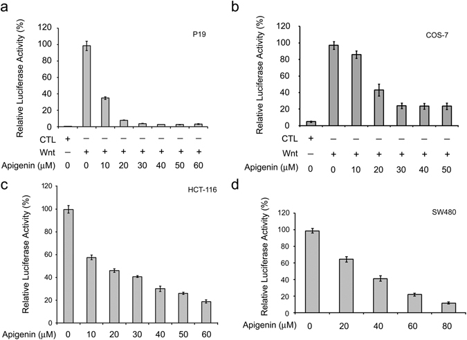
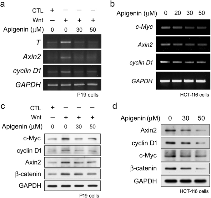
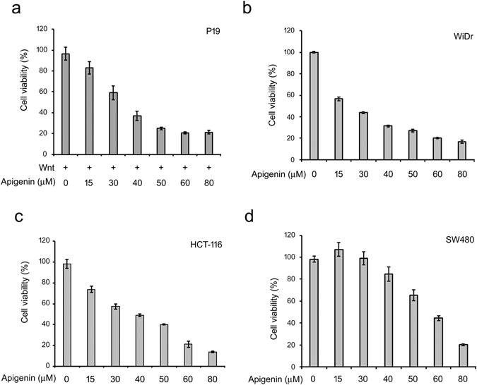
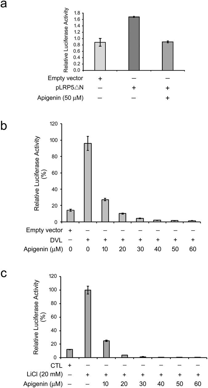
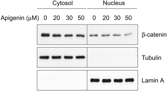
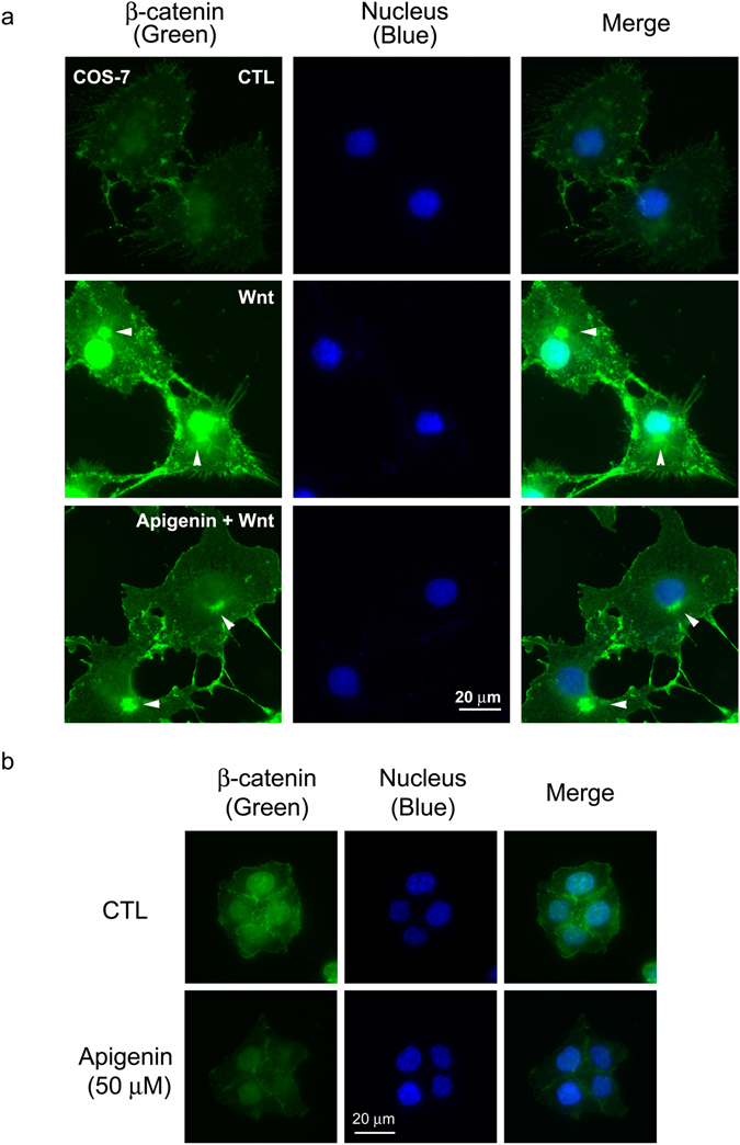
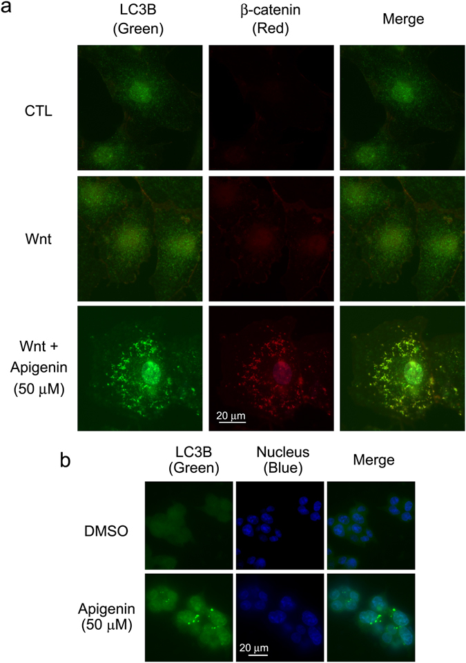
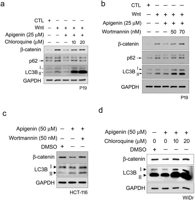
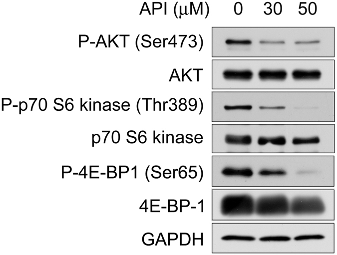
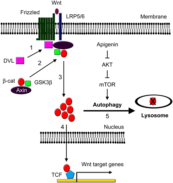
The bioflavonoid apigenin has been shown to possess cancer-preventive and anti-cancer activities. In a drug screening, we found that apigenin can inhibit Wnt/β-catenin signaling, a pathway that participates in pivotal biological functions, which dis-regulation results in various human diseases including cancers. However, the underlying mechanism of apigenin in this pathway and its link to anti-cancer activities remain largely unknown. Here we showed that apigenin reduced the amount of total, cytoplasmic, and nuclear β-catenin, leading to the suppression in the β-catenin/TCF-mediated transcriptional activity, the expression of Wnt target genes, and cell proliferation of Wnt-stimulated P19 cells and Wnt-driven colorectal cancer cells. Western blotting and immunofluorescent staining analyses further revealed that apigenin could induce autophagy-mediated down-regulation of β-catenin in treated cells. Treatment with autophagy inhibitors wortmannin and chloroquine compromised this effect, substantiating the involvement of autophagy-lysosomal system on the degradation of β-catenin during Wnt signaling through inhibition of the AKT/mTOR signaling pathway. Our data not only pointed out a route for the inhibition of canonical Wnt signaling through the induction of autophagy-lysosomal degradation of key player β-catenin, but also suggested that apigenin or other treatments which can initiate this degradation event are potentially used for the therapy of Wnt-related diseases including cancers.


Conflict of interest statement

The authors declare that they have no competing interests.

Figures

Figure 1
Figure 1
Apigenin inhibits Wnt signaling. (a) Apigenin inhibits Wnt signaling of Wnt-stimulated P19 cells. Cells were transfected with the Wnt reporter pGL3-OT and the normalization reporter pTK-Renilla (dual reporters) and treated with control-conditioned medium (CTL), Wnt-3a-conditioned medium (Wnt), or different concentrations (10~60 μM) of apigenin in Wnt-3a-conditioned medium for 16 h, and assayed for dual luciferase activities. (b) Apigenin inhibits Wnt signaling of Wnt-stimulated COS-7 cells. Reporter-transfected cells were treated with control-conditioned medium, Wnt-3a-conditioned medium, or different concentrations (10~50 μM) of apigenin in Wnt-3a-conditioned medium for 20 h, and assayed for dual luciferase activities. Each treatment is in three replicates and data were mean ± S.D. The dual luciferase activity of Wnt-stimulated cells was set as 100% and the relative activity of other treatment was calculated accordingly. (c) Apigenin inhibits intrinsic Wnt signaling in HCT-116 cells. Cells were transfected with dual reporters, treated with different concentrations (0~60 μM) of apigenin for 22 h, and then assayed for dual luciferase activities. (d) Apigenin inhibits intrinsic Wnt signaling in SW480 cells. Reporter-transfected cells were treated with different concentrations (0~80 μM) of apigenin for 22 h, and assayed for dual luciferase activities. Each treatment is in three replicates and data were mean ± S.D. The luciferase activity of treatment without any drug was set as 100%, and the relative activity of other treatment was calculated accordingly.
Figure 2
Figure 2
Apigenin suppresses the expression of Wnt target genes. (a) Apigenin inhibits the mRNA expression of Wnt target genes in Wnt-stimulated P19 cells. Cells were treated with control-conditioned medium, Wnt-3a-conditioned medium, or different concentrations (30, 50 μM) of apigenin in Wnt-3a-conditioned medium for 16 h, and then cells were collected for RT-PCR analyses using gene-specific primers for T, Axin2, cyclin D1, and GAPDH (used as the internal control) as described in methods. (b) Apigenin represses the mRNA expression of Wnt target genes in HCT-116 cells. Cells were treated with different concentrations (0, 20, 30, 50 μM) of apigenin for 22 h and total RNAs were isolated. cDNAs were then synthesized and used for amplification of the specified genes using gene-specific primers for c-Myc, Axin2, cyclin D1, and GAPDH. (c) Apigenin inhibits the protein expression of Wnt target genes in Wnt-stimulated P19 cells. Cells were treated with control-conditioned medium, Wnt-3a-conditioned medium, or different concentrations (30, 50 μM) of apigenin in Wnt-3a-conditioned medium for 16 h, and then cells were collected for Western blotting analyses with anti-c-Myc, anti-cyclin D1, anti-Axin2, anti-beta-catenin, and anti-GAPDH (as the loading control). (d) Apigenin inhibits the protein expression of Wnt target genes in HCT-116 colorectal cancer cells. Cells were treated with different concentrations (0, 30, 50 μM) of apigenin for 22 h and total cell lysates were isolated for Western blotting analyses with anti-Axin2, anti-cyclin D1, anti-c-Myc, anti-β-catenin, and anti-GAPDH.
Figure 3
Figure 3
Apigenin decreases the cell survival of Wnt-stimulated P19 cells and Wnt-driven colorectal cancer cells. (a) Apigenin suppresses the survival of Wnt-stimulated P19 cells. Cells were treated with different concentrations (0~80 μM) of apigenin in Wnt-3a-conditioned medium (Wnt, +) for 4 days and then subjected to MTT assay. The calculated IC50 of apigenin for Wnt-stimulated P19 cells is about 38.15 ± 1.32 μM. (bd) Apigenin suppresses the survival of WiDr, HCT-116 and SW480 cells. Cells were treated with different concentrations (0~80 μM) of apigenin and then subjected to MTT assay. The calculated IC50 of apigenin for WiDr, HCT-116 and SW480 cells is about 29.69 ± 0.15, 39.67 ± 0.99, and 61.38 ± 2.29 μM respectively. Each treatment is in five replicates and data were mean ± S.D.
Figure 4
Figure 4
Apigenin acts on GSK3β per se or downstream of GSK3β in the Wnt signaling pathway. (a) Apigenin acts on LRP5 or downstream of LRP5 in the Wnt/β-catenin signaling pathway. P19 cells were transfected with two reporter plasmids together with pLRP5ΔN (grey bar and heavy grey bar), or empty vector (light grey bar), then treated with 50 μM of apigenin, and assayed for dual luciferase activities. The luciferase activity from the expression of empty vector alone was set as 1.0, and the relative activity of other treatment was calculated accordingly. (b) Apigenin acts on DVL or downstream of DVL in the Wnt signal transduction pathway. P19 cells were transfected with two reporter plasmids together with DVL2 plasmid, or empty vector, then treated with different concentrations (0~60 μM) of apigenin, and assayed for dual luciferase activities. The luciferase activity from the expression of DVL alone was set as 100%, and the relative activity of other treatment was calculated accordingly. (c) Apigenin acts on GSK3β or downstream of GSK3β in the Wnt signaling pathway. P19 cells were transfected with two reporter plasmids, incubated in culture medium (CTL) or treated with 20 mM of LiCl as well as different concentrations (0~60 μM) of apigenin in culture medium, and then assayed for dual luciferase activities. The luciferase activity of LiCl treatment alone was set as 100%, and the relative activity of other treatment was calculated accordingly.
Figure 5
Figure 5
Apigenin decreases the protein levels of cytoplasmic and nuclear β-catenin. HCT-116 cells were treated with different concentrations of apigenin (0, 20, 30, 50 μM) for 22 h. Cells were collected for the isolation of cytosol and nuclear fractions as described in methods. Cytosolic and nuclear lysates were subjected to Western blotting analyses with anti-β-catenin, anti-tubulin, and anti-lamin A. Tubulin served as the cytosolic marker and lamin A served as the nuclear marker.
Figure 6
Figure 6
Apigenin reduces the levels of β-catenin in Wnt-stimulated COS-7 cells and HCT-116 cells by immunofluorescent staining. (a) Apigenin decreases the levels of β-catenin in Wnt-stimulated COS-7 cells. Cells were treated with control-conditioned medium (CTL), Wnt-3a-conditioned medium (Wnt), or 50 μM of apigenin in Wnt-3a-conditioned medium (Apigenin + Wnt) for 20 h, and cells were processed for immunofluorescent staining with β-catenin antibody (labeled in green). Cell nuclei were stained with DAPI (labeled in blue). Arrowheads showed the staining of β-catenin on microtubule-organizing center (MTOC). (b) Apigenin decreases the levels of β-catenin in HCT-116 cells. Cells were either incubated in culture medium (CTL) or treated with 50 μM of apigenin in culture medium for 22 h and processed for immunofluorescent staining with β-catenin antibody (labeled in green). Cell nuclei were stained with DAPI (labeled in blue).
Figure 7
Figure 7
Apigenin induces the formation of autophagosomes in cells. (a) Apigenin induces the formation of autophagosomes in Wnt-stimulated COS-7 cells. COS-7 cells were treated with control-conditioned medium (CTL), Wnt-3a-conditioned medium (Wnt), or 50 μM of apigenin in Wnt-3a-conditioned medium (Wnt + 50 μM Apigenin) for 20 h, and cells were processed for immunofluorescent staining with LC3B antibody (labeled in green) and β-catenin antibody (labeled in red). (b) Apigenin induces the formation autophagosomes in Wnt-driven HCT-116 cells. Cells were treated with carrier reagent (DMSO) or 50 μM of apigenin for 22 h and processed for immunofluorescent staining with LC3B antibody (labeled in green). Cell nuclei were stained with DAPI (labeled in blue).
Figure 8
Figure 8
Autophagy-lysosomal degradation system is involved in apigenin-induced down-regulation of β-catenin during Wnt signaling. Autophagy inhibitor chloroquine (a) and wortmannin (b) inhibit the apigenin-induced degradation of β-catenin upon Wnt stimulation. P19 cells were incubated with control-conditioned medium (CTL), Wnt-3a-conditioned medium (Wnt) or treated with 25 μM of apigenin in Wnt-3a-conditioned medium for 16 h. For autophagy inhibitor treatment, cells were pre-treated with chloroquine (a) and wortmannin (b) for 2 h. Cells were then treated with 25 μM of apigenin and different concentrations of (a) chloroquine (10 and 20 μM) or (b) wortmannin (50 and 70 nM) in Wnt-3a-conditioned medium for 16 h. All cells were collected for Western blotting analyses with β-catenin, p62, LC3B, and GAPDH antibodies. GAPDH served as a loading control. (c) Wortmannin suppresses the apigenin-induced degradation of β-catenin in Wnt-driven HCT-116 cells. Cells were incubated with carrier reagent (DMSO) or 50 μM of apigenin for 22 h. For treatment with autophagy inhibitor, cells were pre-treated with 50 nM of wortmannin for 2 h, and then treated with 50 μM of apigenin and 50 nM of wortmannin for 22 h. All cells were collected for Western blotting analyses with β-catenin, LC3B, and GAPDH antibodies. GAPDH served as a loading control. (d) Chloroquine represses the apigenin-induced degradation of β-catenin in Wnt-driven WiDr cells. Cells were incubated with carrier reagent (DMSO) or 50 μM of apigenin for 22 h. For treatment with autophagy inhibitor, cells were pre-treated with the inhibitor for 2 h. Cells were then treated with 50 μM of apigenin together with different concentrations (10 and 20 μM) of chloroquine for 22 h. All cells were collected for Western blotting analyses with β-catenin, LC3B, and GAPDH antibodies. GAPDH served as a loading control.
Figure 9
Figure 9
Apigenin-mediated down-regulation of β-catenin is through inhibition of the AKT/mTOR signaling pathway. HCT-116 cells were treated with different concentrations of apigenin (0, 30 and 50 μM) for 20 h, and the protein expression of phospho-AKT (Ser473), AKT, phospho-p70 S6 kinase (Thr389), p70 S6 kinase, phospho-4E-BP1 (Ser65), and 4E-BP1 was examined by Western blotting analyses with the antibodies respectively. GAPDH serves as a loading control.
Figure 10
Figure 10
A model depicting the involvement of autophagy-lysosomal system in apigenin-induced degradation of β-catenin during Wnt signaling. In the presence of Wnt, Wnt ligand binds to its cognate receptor Frizzled and co-receptor LRP5/6. Dishevelled (DVL) moves from the cytoplasm to the plasma membrane and associates with Frizzled (step 1). Axin complex, including β-catenin and GSK3β, will also be translocated to the cell membrane and bind to phosphorylated LRP5/6 through Axin (step 2). β-catenin is stabilized and accumulated in the cytoplasm (step 3), and then enters the nucleus to associate with TCF/LEF and activates the expression of Wnt-responsive genes (step 4). Apigenin treatment suppresses the AKT/mTOR signaling pathway and induces the sequestration and degradation of β-catenin inside the lysosome (step 5), contributing to down-regulation of β-catenin during Wnt signaling.

References

    1. Shukla S, Gupta S. Apigenin: a promising molecule for cancer prevention. Pharmaceutical research. 2010;27:962–978. doi: 10.1007/s11095-010-0089-7. - DOI - PMC - PubMed
    1. Srivastava JK, Shankar E, Gupta S. Chamomile: A herbal medicine of the past with bright future. Molecular medicine reports. 2010;3:895–901. - PMC - PubMed
    1. Sarkar FH, Li Y. Cell signaling pathways altered by natural chemopreventive agents. Mutat Res. 2004;555:53–64. doi: 10.1016/j.mrfmmm.2004.04.015. - DOI - PubMed
    1. Arvelo F, Sojo F, Cotte C. Biology of colorectal cancer. Ecancermedicalscience. 2015;9:520. doi: 10.3332/ecancer.2015.520. - DOI - PMC - PubMed
    1. Yang K, et al. The evolving roles of canonical WNT signaling in stem cells and tumorigenesis: implications in targeted cancer therapies. Lab Invest. 2016;96:116–136. doi: 10.1038/labinvest.2015.144. - DOI - PMC - PubMed

Publication types

MeSH terms

LinkOut - more resources