

Use of 3D Printed Bone Plate in Novel Technique to Surgically Correct Hallux Valgus Deformities
- PMID: 28337049
- PMCID: PMC5358518
- DOI: 10.1097/BTO.0000000000000189
Use of 3D Printed Bone Plate in Novel Technique to Surgically Correct Hallux Valgus Deformities
Abstract
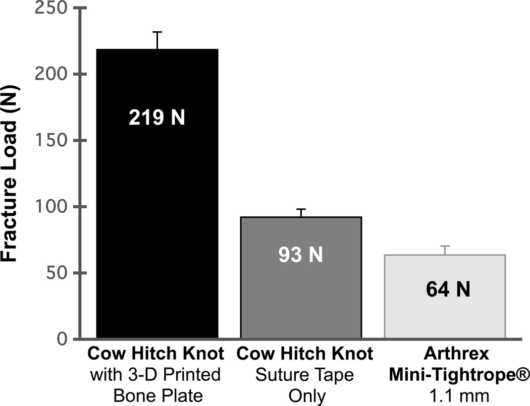
Three-dimensional (3-D) printing offers many potential advantages in designing and manufacturing plating systems for foot and ankle procedures that involve small, geometrically complex bony anatomy. Here, we describe the design and clinical use of a Ti-6Al-4V ELI bone plate (FastForward™ Bone Tether Plate, MedShape, Inc., Atlanta, GA) manufactured through 3-D printing processes. The plate protects the second metatarsal when tethering suture tape between the first and second metatarsals and is a part of a new procedure that corrects hallux valgus (bunion) deformities without relying on doing an osteotomy or fusion procedure. The surgical technique and two clinical cases describing the use of this procedure with the 3-D printed bone plate are presented within.
Keywords: 3-D printing; bone plate; bone tether plate; bunion; fastforward; hallux valgus; metatarsus primus adductus varus; titanium.
Conflict of interest statement
Conflicts of Interest: Dr. Callahan has no financial disclosures or conflicts of interest to report.
Figures


References
-
- Easley ME, Trnka HJ. Current concepts review: hallux valgus part 1: pathomechanics, clinical assessment, and nonoperative management. Foot & ankle international. 2007;28:654–659. - PubMed
-
- Canale ST. Campbell's Operative Orthopaedics. 12th. Mosby: St. Louis; 2010. Disorders of the Hallux; pp. 380–3901.
-
- Easley ME, Trnka HJ. Current concepts review: hallux valgus part II: operative treatment. Foot & ankle international. 2007;28:748–758. - PubMed
-
- Holmes GB, Jr, Hsu AR. Correction of intermetatarsal angle in hallux valgus using small suture button device. Foot & ankle international. 2013;34:543–549. - PubMed
-
- Weatherall JM, Chapman CB, Shapiro SL. Postoperative second metatarsal fractures associated with suture-button implant in hallux valgus surgery. Foot & ankle international. 2013;34:104–110. - PubMed
Grants and funding
LinkOut - more resources
Full Text Sources
Other Literature Sources