

Late spontaneous resolution of a double anterior chamber post deep anterior lamellar keratoplasty
- PMID: 28337067
- PMCID: PMC5352942
- DOI: 10.1016/j.sjopt.2017.01.003
Late spontaneous resolution of a double anterior chamber post deep anterior lamellar keratoplasty
Abstract
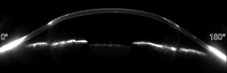
A 31-year-old healthy male underwent deep anterior lamellar keratoplasty with big-bubble technique for treatment of keratoconus in his right eye. One week after surgery, he presented with detachment of the endothelium-Descemet complex with formation of a double anterior chamber, despite the apparent absence of an intraoperative Descemet membrane rupture. A subsequent intervention with the intent to relocate the corneal graft button was not effective, because the detachment appeared again one day later. The authors hypothesized that, at the time of the stromal dissection with big bubble technique, a small amount of air penetrated into the anterior chamber, creating a false pathway through the trabecular meshwork. The aqueous humor then penetrated the graft flowing through the false pathway, causing the endothelium-Descemet detachment. The persistence of that pathway, even after the intervention of graft repositioning, caused the failure of the latter procedure and persistence of the double chamber. We decided to wait and observe. The double anterior chamber spontaneously resolved in approximately three months.
Keywords: Big-bubble technique; C3F8, perfluoropropane; Deep anterior lamellar keratoplasty; Double anterior chamber; Keratoconus; SF6, sulfur hexafluoride.
Figures


References
-
- Anwar M., Teichmann K.D. Big-bubble technique to bare Descemet's membrane in anterior lamellar keratoplasty. J Cataract Refractive Surg. 2002;28(3):398–403. - PubMed
-
- Han D.C., Mehta J.S., Por Y.M., Htoon H.M., Tan D.T. Comparison of outcomes of lamellar keratoplasty and penetrating keratoplasty in keratoconus. Am J Ophthalmology. 2009;148(5):744–751. 744–51.e1. - PubMed
-
- Leccisotti A. Descemet's membrane perforation during deep anterior lamellar keratoplasty: prognosis. J Cataract Refractive Surg. 2007;33(5):825–829. - PubMed
-
- Jhanji V., Sharma N., Vajpayee R.B. Intraoperative perforation of Descemet's membrane during “big bubble” deep anterior lamellar keratoplasty. Int Ophthalmology. 2010;30(3):291–295. - PubMed
LinkOut - more resources
Full Text Sources
Other Literature Sources
