

Peritoneal tuberculosis with elevated CA-125 mimicking ovarian cancer with carcinomatosis peritonei: Crucial CT findings
- PMID: 28337102
- PMCID: PMC5318682
- DOI: 10.17179/excli2016-625
Peritoneal tuberculosis with elevated CA-125 mimicking ovarian cancer with carcinomatosis peritonei: Crucial CT findings
Abstract
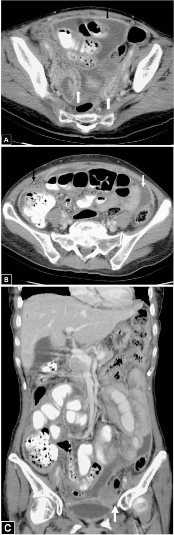
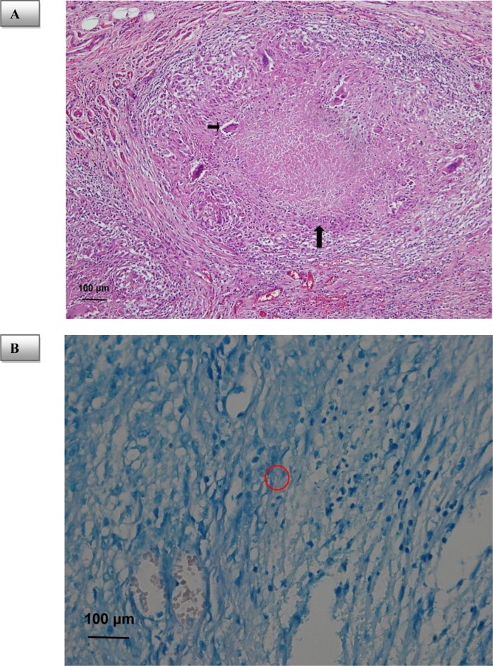
Preoperative diagnosis of peritoneal tuberculosis is often difficult because of confusion with ovarian cancer. A 56-year-old woman was admitted to our hospital with abdominal fullness. Ascites, prominent bilateral ovaries, and elevated CA-125 were noted. Computed tomography showed thickened peritoneum and strandings in the mesentery and omentum. Exploratory laparotomy was performed under the provisional diagnosis of ovarian cancer, but the final diagnosis was peritoneal tuberculosis. Careful evaluation of bilateral fallopian tubes and ovaries and peritoneum are helpful for correct diagnosis.
Keywords: carcinomatosis peritonei; computed tomography (CT); ovarian cancer; peritoneum; tuberculosis.
Figures
Similar articles
-
Tuberculous peritonitis mimicking carcinomatosis peritonei: CT findings and histopathologic correlation.Radiol Case Rep. 2019 Oct 16;14(12):1491-1494. doi: 10.1016/j.radcr.2019.09.026. eCollection 2019 Dec. Radiol Case Rep. 2019. PMID: 31660094 Free PMC article.
-
An unusual case of dedifferentiated leiomyosarcoma of the primary mesentery mimicking ovarian cancer.Int J Clin Exp Pathol. 2019 Nov 1;12(11):4150-4155. eCollection 2019. Int J Clin Exp Pathol. 2019. PMID: 31933813 Free PMC article.
-
Peritoneal Tuberculosis Mimicking Ovarian Malignancy: A Case Report.Cureus. 2024 Dec 22;16(12):e76231. doi: 10.7759/cureus.76231. eCollection 2024 Dec. Cureus. 2024. PMID: 39845238 Free PMC article.
-
US of the peritoneum.Radiographics. 2003 May-Jun;23(3):663-84; discussion 684-5. doi: 10.1148/rg.233025712. Radiographics. 2003. PMID: 12740467 Review.
-
CT imaging of peritoneal carcinomatosis and its mimics.Diagn Interv Imaging. 2014 Sep;95(9):861-72. doi: 10.1016/j.diii.2014.02.009. Epub 2014 Mar 14. Diagn Interv Imaging. 2014. PMID: 24631039 Review.
Cited by
-
A diagnostic approach for differentiating abdominal tuberculosis from ovarian malignancy: a case series and literature review.BMC Proc. 2019 Dec 16;13(Suppl 11):13. doi: 10.1186/s12919-019-0180-y. eCollection 2019. BMC Proc. 2019. PMID: 31890006 Free PMC article.
-
Ovarian mass - tuberculosis or malignancy? Need for early intensified evaluation.EXCLI J. 2018 Sep 14;17:914-915. doi: 10.17179/excli2018-1640. eCollection 2018. EXCLI J. 2018. PMID: 30564070 Free PMC article. No abstract available.
-
Expression of Circulating MicroRNA-141 in Epithelial Ovarian Cancer.Malays J Med Sci. 2020 Dec;27(6):27-38. doi: 10.21315/mjms2020.27.6.4. Epub 2020 Dec 1. Malays J Med Sci. 2020. PMID: 33447132 Free PMC article.
-
Diagnostic value of serum CA125 combined with PET/CT in ovarian cancer and tuberculous peritonitis in female patients.Abdom Radiol (NY). 2023 Nov;48(11):3449-3457. doi: 10.1007/s00261-023-03997-9. Epub 2023 Jul 26. Abdom Radiol (NY). 2023. PMID: 37493838
References
-
- Barutcu O, Erel HE. Abdominopelvic tuberculosis simulating disseminated ovarian carcinoma with elevated CA-125 level: report of two cases. Abdom Imag. 2002;27:465–470. - PubMed
-
- Bilgin T, Karabay A, Dolar E, Develioğlu OH. Peritoneal tuberculosis with pelvic abdominal mass, ascites and elevated CA 125 mimicking advanced ovarian carcinoma: a series of 10 cases. Int J Gynecol Cancer. 2001;11:290–294. - PubMed
-
- Chow KM, Chow VC, Hung LC, Wong SM, Szeto CC. Tuberculous peritonitis-associated mortality is high among patients waiting for the results of mycobacterial cultures of ascites fluid samples. Clin Infect Dis. 2002;35:409–413. - PubMed
-
- Koc S, Beydilli G, Tulunay G, Ocalan R, Boran N, Ozgul N, et al. Peritoneal tuberculosis mimicking advanced ovarian cancer: a retrospective review of 22 cases. Gynecol Oncol. 2006;103:565–569. - PubMed
-
- Leder RA, Low VHS. Tuberculosis of the abdomen. Radiol Clin North Am. 1995;33:691–705. - PubMed
Publication types
LinkOut - more resources
Full Text Sources
Research Materials
Miscellaneous