Review
. 2017:2017:8482326.
doi: 10.1155/2017/8482326. Epub 2017 Feb 28.

Gut Mesenchymal Stromal Cells in Immunity

Affiliations
Review

Gut Mesenchymal Stromal Cells in Immunity

Valeria Messina et al. Stem Cells Int. 2017.

Abstract

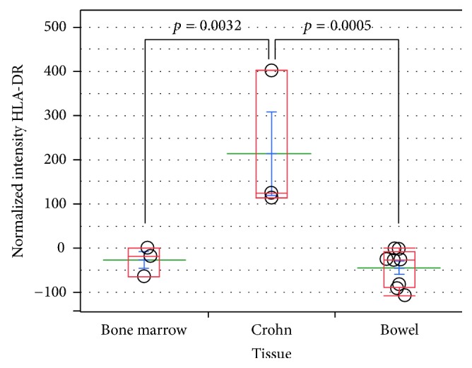
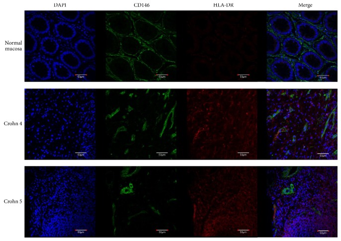
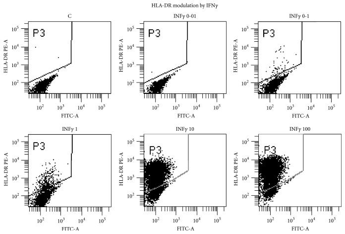
Mesenchymal stromal cells (MSCs), first found in bone marrow (BM), are the structural architects of all organs, participating in most biological functions. MSCs possess tissue-specific signatures that allow their discrimination according to their origin and location. Among their multiple functions, MSCs closely interact with immune cells, orchestrating their activity to maintain overall homeostasis. The phenotype of tissue MSCs residing in the bowel overlaps with myofibroblasts, lining the bottom walls of intestinal crypts (pericryptal) or interspersed within intestinal submucosa (intercryptal). In Crohn's disease, intestinal MSCs are tightly stacked in a chronic inflammatory milieu, which causes their enforced expression of Class II major histocompatibility complex (MHC). The absence of Class II MHC is a hallmark for immune-modulator and tolerogenic properties of normal MSCs and, vice versa, the expression of HLA-DR is peculiar to antigen presenting cells, that is, immune-activator cells. Interferon gamma (IFNγ) is responsible for induction of Class II MHC expression on intestinal MSCs. The reversal of myofibroblasts/MSCs from an immune-modulator to an activator phenotype in Crohn's disease results in the formation of a fibrotic tube subverting the intestinal structure. Epithelial metaplastic areas in this context can progress to dysplasia and cancer.


Conflict of interest statement

The authors declare that they have no competing interests.

Figures

Figure 1
Figure 1
HLA-DR levels in Crohn's-derived versus bowel- and bone marrow-derived MSCs. Dot plot of normalized fluorescence intensities from flow cytometric analysis of HLA-DR expression in Crohn, colon, and bone marrow MSCs. MSCs obtained from colon and bone marrow samples are consistently negative for HLA-DR, a major histocompatibility complex (MHC) Class II antigen, whereas Crohn's-derived MSCs constitutively express statistically significant levels. The antibody used was R-phycoerythrin-labeled mouse anti-human HLA-DR (Becton-Dickinson). Mean fluorescence intensity (MFI) of isotype control was subtracted to MFI from HLA-DR intensity to generate the plotted normalized intensities. p values were obtained by performing Student's t-test via SAS JMP v10 software (SAS Institute Inc., Cary, NC).
Figure 2
Figure 2
Immunofluorescence analysis of Crohn's disease surgical specimens. In contrast to the typical structure of normal distal intestine mucosa, Crohn's disease sections have a totally subverted architecture wherein the presence of some CD146+ cells expressing high levels of HLA-DR (white arrows) is detected, in close proximity to or interspersed between smaller HLA-DR+ cells. Normal colonic mucosa and Crohn's disease specimens were obtained from patients who signed an informed consent before undergoing surgical resection. Tissues were fixed in PFA 4% o/n at +4°C, embedded, frozen in OCT, and stored at −80°C. Thick sections (15 μm) were cut on a cryotome and processed for immunofluorescence staining. Permeabilization was performed with 0.5% Triton X-100 in PBS for 30′, at room temperature. A blocking step was performed using 40 μg/mL mouse IgG in 3% BSA (PBS) for 2 h at RT, and subsequent incubation with primary antibodies was carried out for 16h at +4°C. Primary antibodies used were FITC conjugated mouse anti-CD146 (Biocytex) and R phycoerythrin-labeled mouse anti-human HLA-DR (Becton-Dickinson). The final step, before mounting the slides, was incubation with a TRITC-conjugated goat anti-PE secondary antibody (Aviva Systems Biology), for 2 h at 37°C. Nuclear staining was executed adding RNAse and DAPI to the secondary antibody incubation mix. Images were acquired with an Olympus FV-1000 spectral confocal microscope, equipped with an UPLFLN 40x 1.30 NA oil immersion objective. Scale bars correspond to 50 μm.
Figure 3
Figure 3
Dot plot analysis of C-MSCs treated with graded amount of IFNγ ng/ml for 72 h. Bowel MSCs were stained with PE-labeled HLA-DR mAB (Pharmingen) and analysed with FACSCanto (Becton-Dickinson).

References

    1. Sorrentino A., Ferracin M., Castelli G., et al. Isolation and characterization of CD146+ multipotent mesenchymal stromal cells. Experimental Hematology. 2008;36(8):1035–1046. doi: 10.1016/j.exphem.2008.03.004. - DOI - PubMed
    1. Valtieri M., Sorrentino A. The mesenchymal stromal cell contribution to homeostasis. Journal of Cellular Physiology. 2008;217(2):296–300. doi: 10.1002/jcp.21521. - DOI - PubMed
    1. Signore M., Cerio A. M., Boe A., et al. Identity and ranking of colonic mesenchymal stromal cells. Journal of Cellular Physiology. 2012;227(9):3291–3300. doi: 10.1002/jcp.24027. - DOI - PubMed
    1. Picchi J., Trombi L., Spugnesi L., et al. HOX and TALE signatures specify human stromal stem cell populations from different sources. Journal of Cellular Physiology. 2013;228(4):879–889. doi: 10.1002/jcp.24239. - DOI - PubMed
    1. Parekkadan B., Tilles A. W., Yarmush M. L. Bone marrow-derived mesenchymal stem cells ameliorate autoimmune enteropathy independently of regulatory T cells. Stem Cells. 2008;26(7):1913–1919. doi: 10.1634/stemcells.2007-0790. - DOI - PubMed