

Differential involvement of corticospinal tract (CST) fibers in UMN-predominant ALS patients with or without CST hyperintensity: A diffusion tensor tractography study
- PMID: 28337412
- PMCID: PMC5349615
- DOI: 10.1016/j.nicl.2017.02.017
Differential involvement of corticospinal tract (CST) fibers in UMN-predominant ALS patients with or without CST hyperintensity: A diffusion tensor tractography study
Abstract
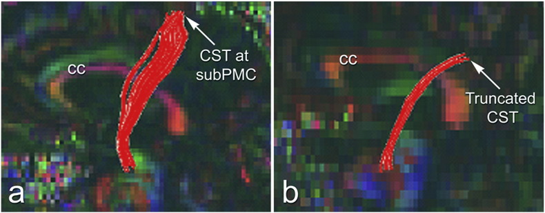
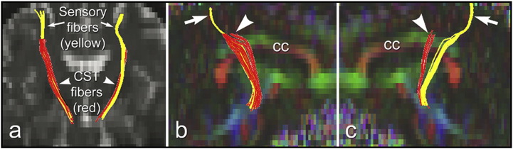
Diagnosis of amyotrophic lateral sclerosis (ALS) depends on clinical evidence of combined upper motor neuron (UMN) and lower motor neuron (LMN) degeneration, although ALS patients can present with features predominantly of one or the other. Some UMN-predominant patients show hyperintense signal along the intracranial corticospinal tract (CST) on T2- and proton density (PD)-weighted images (ALS-CST +), and appear to have faster disease progression when compared to those without CST hyperintensity (ALS-CST -). The reason for this is unknown. We hypothesized that diffusion tensor tractography (DTT) would reveal differences in DTI abnormalities along the intracranial CST between these two patient subgroups. Clinical DTI scans were obtained at 1.5T in 14 neurologic controls and 45 ALS patients categorized into two UMN phenotypes based on clinical measures and MRI. DTT was used to quantitatively assess the CST in control and ALS groups. DTT revealed subcortical loss ('truncation') of virtual motor CST fibers (presumably) projecting from the precentral gyrus (PrG) in ALS patients but not in controls; in contrast, virtual fibers (presumably) projecting to the adjacent postcentral gyrus (PoG) were spared. No significant differences in virtual CST fiber length were observed between controls and ALS patients. However, the frequency of CST truncation was significantly higher in the ALS-CST + subgroup (9 of 21) than in the ALS-CST - subgroup (4 of 24; p = 0.049), suggesting this finding could differentiate these ALS subgroups. Also, because virtual CST truncation occurred only in the ALS patient group and not in the control group (p = 0.018), this DTT finding could prove to be a diagnostic biomarker of ALS. Significantly shorter disease duration and faster disease progression rate were observed in ALS patients with CST fiber truncation than in those without (p < 0.05). DTI metrics of fractional anisotropy (FA), mean diffusivity (MD), axial diffusivity (AD) and radial diffusivity (RD) were also determined in four regions of interest (ROIs) along the CST, namely: cerebral peduncle (CP), posterior limb of internal capsule (PLIC), centrum semiovale at top of lateral ventricle (CSoLV) and subcortical to primary motor cortex (subPMC). Of note, FA values along the left hemisphere virtual CST tract were significantly different between controls and ALS-CST + patients (p < 0.05) only at the PLIC level, but not at the CSoLV or subPMC level. Also, no significant differences in FA values were observed between ALS subgroups or between control and ALS-CST - groups (p > 0.05) in any of the ROIs. In addition, comparing FA values between ALS patients with CST truncation and those without in the aforementioned four ROIs, revealed no significant differences in either hemisphere. However, visual evaluation of DTT was able to identify UMN degeneration in patients with ALS, particularly in those with a more aggressive clinical disease course and possibly different pathologic processes.
Keywords: ALS; ALS, Amyotrophic lateral sclerosis; CNS, Central nervous system; CP, Cerebral peduncle; CST, Corticospinal tract; CSoLV, Centrum semiovale at top of lateral ventricle; DTI; DTI, Diffusion tensor imaging; DTT, Diffusion tensor tractography; DW, Diffusion weighted; Diffusion tensor tractography; EMG, Electromyography; EPI, Echo planar imaging; FA, Fractional anisotropy; FLAIR, Fluid attenuated inversion recovery; FSE, Fast spin echo; LMN, Lower motor neuron; MD, Mean diffusivity; MR, Magnetic resonance; MRI, Magnetic resonance imaging; PD, Proton density; PLIC, Posterior limb of the internal capsule; PMC, Primary motor cortex; PSC, Primary sensory cortex; Phenotypes; PoG, Postcentral gyrus; PrG, Precentral gyrus; ROI, Region of interest; SNR, Signal-to-noise ratio; SS-EPI, Single shot echo planar imaging; SubPMC, Subcortical to primary motor cortex; TE, Echo time; TR, Repetition time; UMN, Upper motor neuron; cMRI, Conventional MRI.
Figures
References
-
- da Rocha A.J., Oliveira A.S., Fonseca R.B., Maia A.C., Jr., Buainain R.P., Lederman H.M. Detection of corticospinal tract compromise in amyotrophic lateral sclerosis with brain MR imaging: relevance of the T1-weighted spin-echo magnetization transfer contrast sequence. AJNR Am. J. Neuroradiol. 2004 Oct;25(9):1509–1515. (PubMed PMID: 15502129. Epub 2004/10/27. eng) - PMC - PubMed
MeSH terms
LinkOut - more resources
Full Text Sources
Other Literature Sources
Medical
Research Materials
Miscellaneous
