Review
. 2017:2017:9217145.
doi: 10.1155/2017/9217145. Epub 2017 Feb 26.

Caudal Epidural Block: An Updated Review of Anatomy and Techniques

Affiliations
Review

Caudal Epidural Block: An Updated Review of Anatomy and Techniques

Sheng-Chin Kao et al. Biomed Res Int. 2017.

Abstract

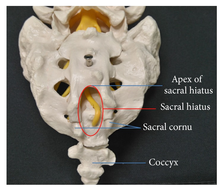
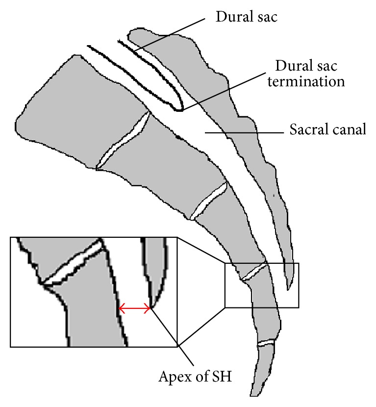
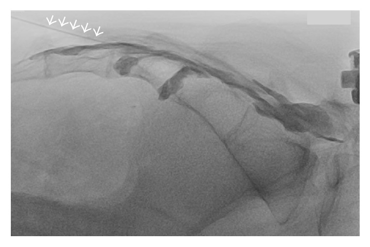
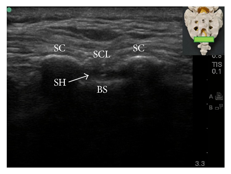
Caudal epidural block is a commonly used technique for surgical anesthesia in children and chronic pain management in adults. It is performed by inserting a needle through the sacral hiatus to gain entrance into the sacral epidural space. Using conventional blind technique, the failure rate of caudal epidural block in adults is high even in experienced hands. This high failure rate could be attributed to anatomic variations that make locating sacral hiatus difficult. With the advent of fluoroscopy and ultrasound in guiding needle placement, the success rate of caudal epidural block has been markedly improved. Although fluoroscopy is still considered the gold standard when performing caudal epidural injection, ultrasonography has been demonstrated to be highly effective in accurately guiding the needle entering the caudal epidural space and produce comparative treatment outcome as fluoroscopy. Except intravascular and intrathecal injection, ultrasonography could be as effective as fluoroscopy in preventing complications during caudal epidural injection. The relevant anatomy and techniques in performing the caudal epidural block will be briefly reviewed in this article.


Conflict of interest statement

The authors declare that there is no conflict of interests regarding the publication of this paper.

Figures

Figure 1
Figure 1
Posterior view of sacrum.
Figure 2
Figure 2
Sagittal view of sacrum. SH: sacral hiatus; red double-ended arrow: anterior-posterior diameter of sacral hiatus at its apex.
Figure 3
Figure 3
Fluoroscopy-guided caudal epidural block. Proper needle tip placement was verified by observing spread of contrast medium within the epidural space without intravascular uptake. Arrows: needle.
Figure 4
Figure 4
Transverse ultrasound view of the sacral hiatus. The inset shows the position of the ultrasound transducer. BS: base of sacrum; SC: sacral cornua; SCL: sacrococcygeal ligament; SH: sacral hiatus.
Figure 5
Figure 5
Longitudinal ultrasound view of sacral hiatus. The inset shows the position of the ultrasound transducer. BS: base of sacrum; SCL: sacrococcygeal ligament; SH: sacral hiatus; arrows: needle.
Figure 6
Figure 6
Color Doppler ultrasonography in longitudinal view of sacral hiatus. A predominantly one-color spectrum is observed in the sacral hiatus during caudal epidural injection. The inset shows the position of the ultrasound transducer. BS: base of sacrum; SCL: sacrococcygeal ligament; SH: sacral hiatus.
Figure 7
Figure 7
Color Doppler ultrasonography in paramedian sagittal oblique view of the sacral and lumbar spine. The observed color spectrum suggests the flow of injectate reaching L4-5 level. The inset shows the position of the ultrasound transducer. L3L: L3 lamina; L4L: L4 lamina; L5L: L5 lamina; DS: dorsal surface of sacrum.

Similar articles

Cited by

References

    1. Dalens B., Hasnaoui A. Caudal anesthesia in pediatric surgery: success rate and adverse effects in 750 consecutive patients. Anesthesia and Analgesia. 1989;68(2):83–89. doi: 10.1213/00000539-198902000-00002. - DOI - PubMed
    1. Orme R. M., Berg S. J. The ‘swoosh’ test—an evaluation of a modified ‘whoosh’ test in children. British Journal of Anaesthesia. 2003;90:62–65. - PubMed
    1. Renfrew D. L., Moore T. E., Kathol M. H., El-Khoury G. Y., Lemke J. H., Walker C. W. Correct placement of epidural steroid injections: fluoroscopic guidance and contrast administration. American Journal of Neuroradiology. 1991;12(5):1003–1007. - PMC - PubMed
    1. Stitz M. Y., Sommer H. M. Accuracy of blind versus fluoroscopically guided caudal epidural injection. Spine. 1999;24(13):1371–1376. doi: 10.1097/00007632-199907010-00016. - DOI - PubMed
    1. Barham G., Hilton A. Caudal epidurals: the accuracy of blind needle placement and the value of a confirmatory epidurogram. European Spine Journal. 2010;19(9):1479–1483. doi: 10.1007/s00586-010-1469-8. - DOI - PMC - PubMed

LinkOut - more resources