

The impact of mangiferin from Belamcanda chinensis on experimental colitis in rats
- PMID: 28337639
- PMCID: PMC5859701
- DOI: 10.1007/s10787-017-0337-0
The impact of mangiferin from Belamcanda chinensis on experimental colitis in rats
Abstract
Background: Inflammatory bowel disease (IBD) [including Crohn's disease (CD) and ulcerative colitis (UC)] constitutes an important clinical problem. The pathogenesis of IBD remains unclear. It is believed that immune dysfunction, inflammatory mediators and oxidative damage play crucial roles in development of IBD. The condition is clinically associated with symptoms ranging from mild to severe during relapses, depending on the affected segment of the gastrointestinal tract. Bloody diarrhea with mucus, abdominal pain, weight loss and anemia are initial symptoms of both CD and UC. Differences between diseases become more evident in time, along with the development of intestinal and extraintestinal complications. Mangiferin (1,3,6,7-tetrahydroxyxanthone-C-2-β-D-glucoside), a natural polyphenol in plants, exerts antioxidant and anti-inflammatory effects making it an interesting option for the treatment of inflammatory pathologies associated with oxidative stress in humans, such as IBD.
Purpose: The aim of the current study was to elucidate the impact of mangiferin on colon tissues in 2,4,6-trinitrobenzensulfonic acid (TNBS)-induced colitis in rats.
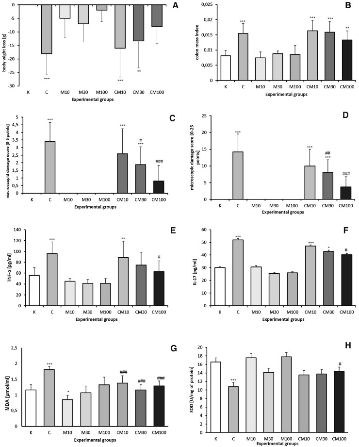
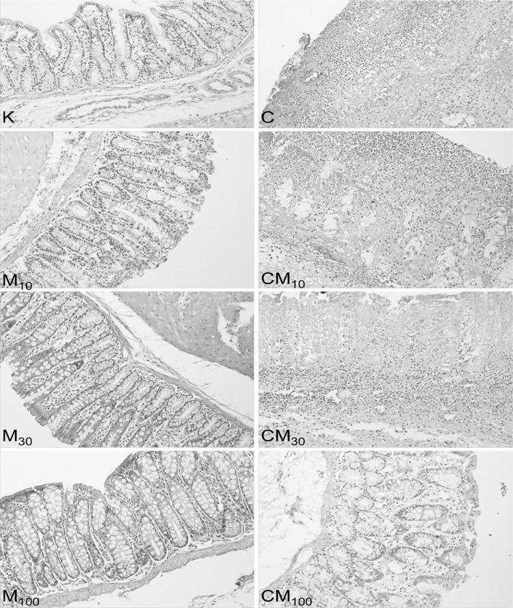
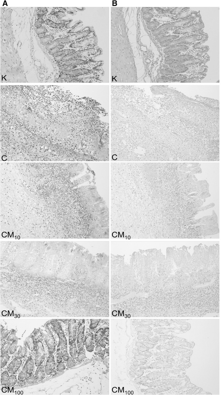
Methods: Mangiferin was obtained from Belamcanda chinensis rhizomes by a multistage process. Groups of rats were pre-treated with 10, 30 or 100 mg/kg of mangiferin, or with distilled water administered intragastrically for 16 days. An ethanol solution of TNBS or saline was given rectally on the day 15 of the experiment. The experiment was terminated on the day 17. The colon was removed, cleaned, weighed and examined macro- and microscopically. Determination of tumor necrosis factor α (TNF-α), interleukin 17 (IL-17), malondialdehyde (MDA) levels and superoxide dismutase (SOD) activity were performed spectrophotometrically in homogenates of colon tissues.
Results: Rats in the TNBS group developed symptoms of colitis, including: body weight loss, colon mass index increase and damage of intestinal tissues with concomitant increase in TNF-α, IL-17, MDA levels and decreased SOD activity. In non-TNBS-treated rats mangiferin did not cause any changes of studied parameters. Pre-treatment with mangiferin exerted a protective effect, reducing the intensity of damage caused by TNBS. Mangiferin at the doses of 30 and 100 mg/kg reduced the macro- and microscopic damage score and the MDA level in colon tissues. Only at the dose of 100 mg/kg, mangiferin decreased TNF-α and IL-17 concentrations, and SOD activity in colon tissues.
Conclusion: Mangiferin attenuates inflammatory changes of colon tissues in experimental, TNBS-induced colitis in rats. Protective effect exerted by mangiferin depends primarily on its anti-inflammatory activity and secondarily on its antioxidant properties.
Keywords: Experimental colitis; Inflammatory bowel disease; Interleukin-17; Mangiferin; Trinitrobenzensulfonic acid; Tumor necrosis factor α.
Conflict of interest statement
None.
Figures

Similar articles
-
Copaifera malmei Harms leaves infusion attenuates TNBS-ulcerative colitis through modulation of cytokines, oxidative stress and mucus in experimental rats.J Ethnopharmacol. 2021 Mar 1;267:113499. doi: 10.1016/j.jep.2020.113499. Epub 2020 Oct 19. J Ethnopharmacol. 2021. PMID: 33091486
-
Mangiferin attenuates DSS colitis in mice: Molecular docking and in vivo approach.Chem Biol Interact. 2016 Jun 25;253:18-26. doi: 10.1016/j.cbi.2016.04.033. Epub 2016 Apr 26. Chem Biol Interact. 2016. PMID: 27125760
-
Protective effect of Averrhoa bilimbi L. fruit extract on ulcerative colitis in wistar rats via regulation of inflammatory mediators and cytokines.Biomed Pharmacother. 2017 Jul;91:1113-1121. doi: 10.1016/j.biopha.2017.05.057. Epub 2017 May 16. Biomed Pharmacother. 2017. PMID: 28531922
-
Cytokines in colitis associated cancer: potential drug targets?Inflamm Allergy Drug Targets. 2008 Sep;7(3):187-94. doi: 10.2174/187152808785748137. Inflamm Allergy Drug Targets. 2008. PMID: 18782026 Review.
-
Saffron as a Promising Therapy for Inflammatory Bowel Disease.Nutrients. 2024 Jul 20;16(14):2353. doi: 10.3390/nu16142353. Nutrients. 2024. PMID: 39064796 Free PMC article. Review.
Cited by
-
Soluble Protein Hydrolysate Ameliorates Gastrointestinal Inflammation and Injury in 2,4,6-Trinitrobenzene Sulfonic Acid-Induced Colitis in Mice.Biomolecules. 2022 Sep 13;12(9):1287. doi: 10.3390/biom12091287. Biomolecules. 2022. PMID: 36139127 Free PMC article.
-
Inducing Apoptosis and Suppressing Inflammatory Reactions in Synovial Fibroblasts are Two Important Ways for Guizhi-Shaoyao-Zhimu Decoction Against Rheumatoid Arthritis.J Inflamm Res. 2021 Jan 26;14:217-236. doi: 10.2147/JIR.S287242. eCollection 2021. J Inflamm Res. 2021. PMID: 33542641 Free PMC article.
-
From Nature to Nanotechnology: The Bioactivities of Mangiferin Explored.Nanotechnol Sci Appl. 2025 Jul 10;18:277-294. doi: 10.2147/NSA.S525423. eCollection 2025. Nanotechnol Sci Appl. 2025. PMID: 40662091 Free PMC article. Review.
-
Pharmacokinetic and metabolomic analyses of Mangiferin calcium salt in rat models of type 2 diabetes and non-alcoholic fatty liver disease.BMC Pharmacol Toxicol. 2020 Aug 6;21(1):59. doi: 10.1186/s40360-020-00438-x. BMC Pharmacol Toxicol. 2020. PMID: 32762728 Free PMC article.
-
Supercritical Fluid Extract of Angelica sinensis and Zingiber officinale Roscoe Ameliorates TNBS-Induced Colitis in Rats.Int J Mol Sci. 2019 Aug 5;20(15):3816. doi: 10.3390/ijms20153816. Int J Mol Sci. 2019. PMID: 31387229 Free PMC article.
References
-
- Arribas B, Suárez-Pereira E, Ortiz-Mellet C, García-Fernández JM, Buttersack C, Rodríguez-Cabezas ME, et al. Di-d-fructose dianhydride-enriched caramels: effect on colon microbiota, inflammation, and tissue damage in trinitrobenzensulfonic acid-induced colitic rats. J Agric Food Chem. 2010;58:6476–6484. doi: 10.1021/jf100513j. - DOI - PubMed
-
- Dignass A, Lindsay JO, Sturm A, Windsor A, Colombel JF, Allez M, et al. Second European evidence-based consensus on the diagnosis and management of ulcerative colitis. Part 2: current management. JCC. 2012;6:991–1030. - PubMed
MeSH terms
Substances
Grants and funding
LinkOut - more resources
Full Text Sources
Other Literature Sources