Review
. 2017 May;90(1073):20160783.
doi: 10.1259/bjr.20160783. Epub 2017 Mar 24.

Acquired pulmonary artery pseudoaneurysms: a pictorial review

Affiliations
Review

Acquired pulmonary artery pseudoaneurysms: a pictorial review

Benedicte Guillaume et al. Br J Radiol. 2017 May.

Abstract

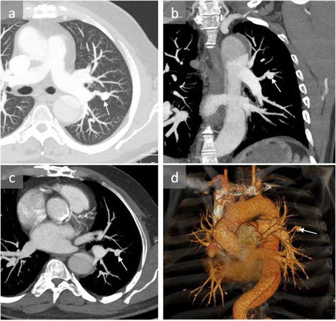
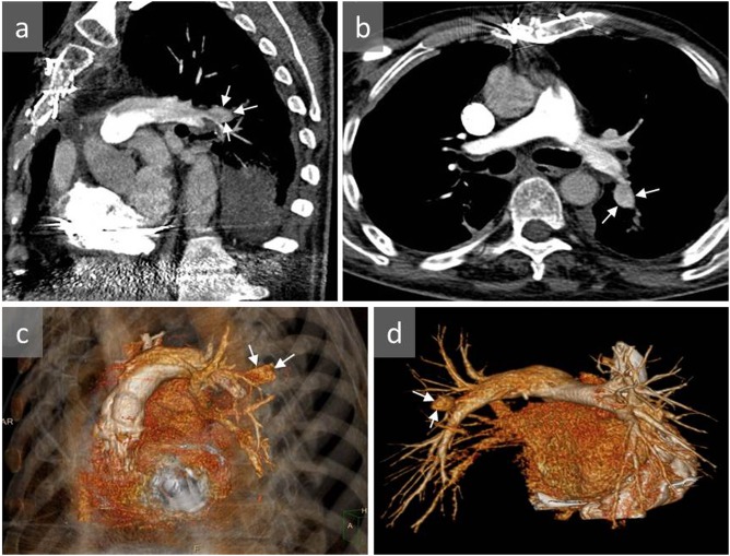
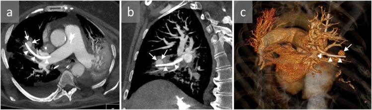
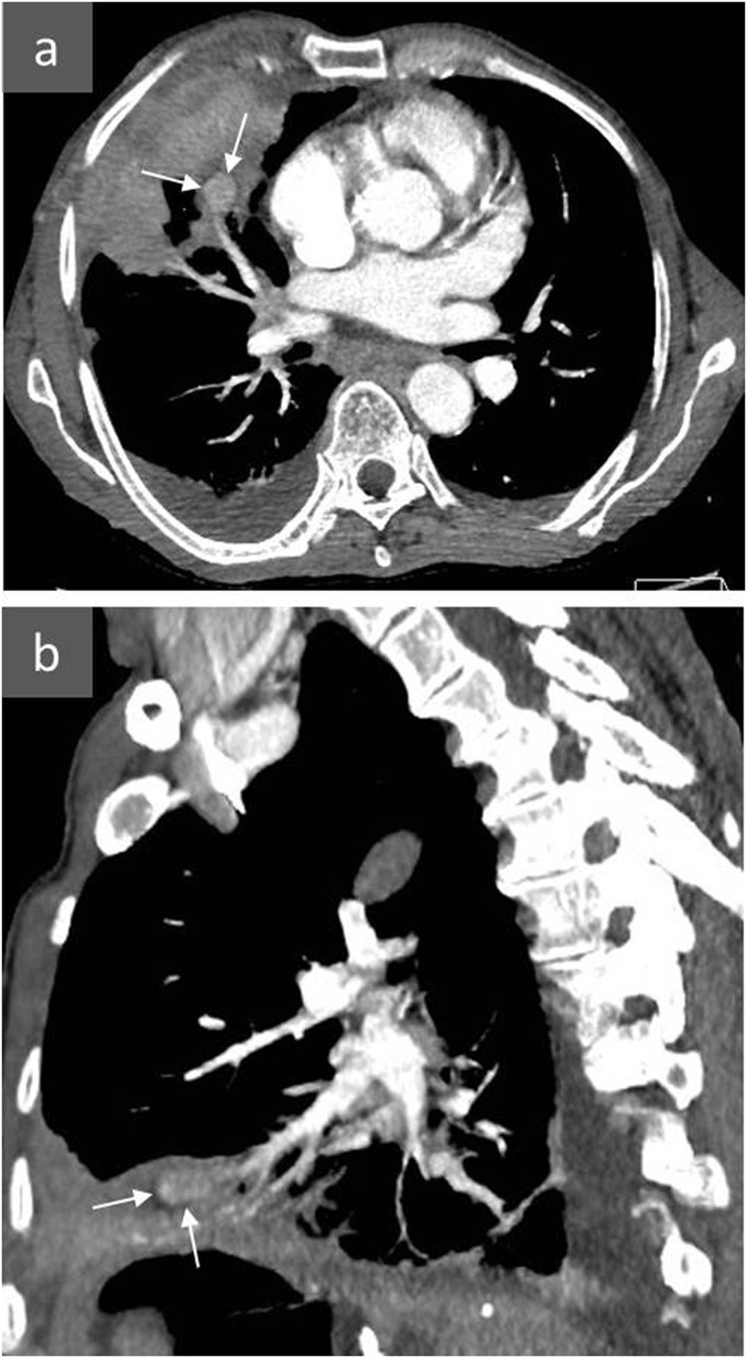
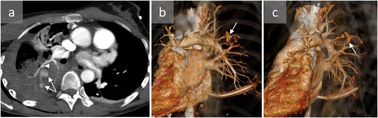
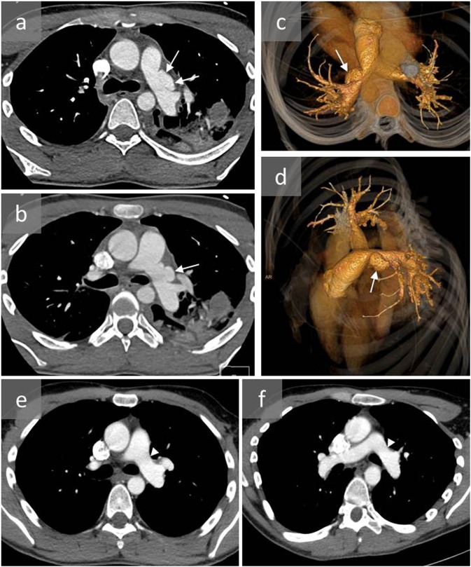
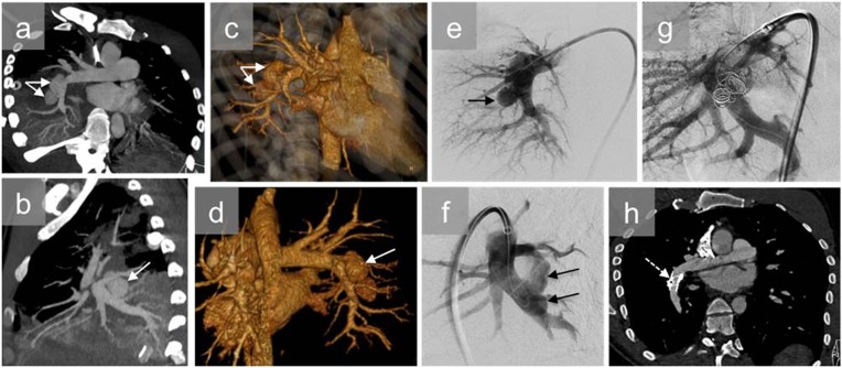
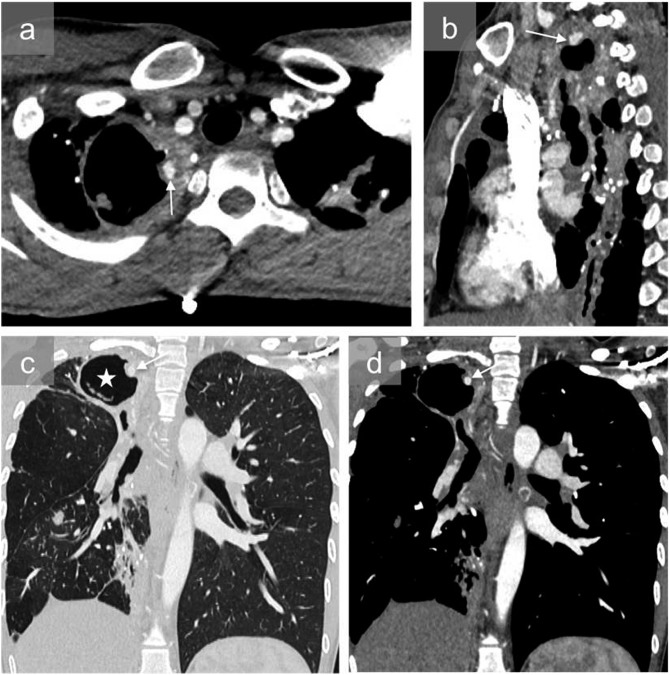
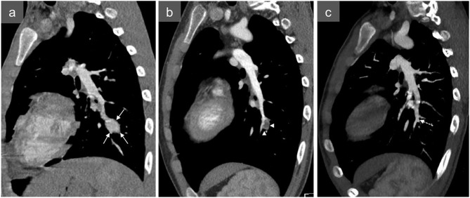
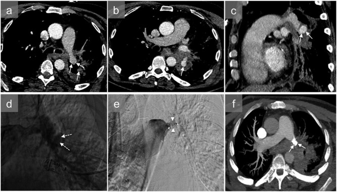
Pulmonary artery pseudoaneurysms (PAPs) are uncommon but potentially lethal. They may be incidentally discovered on imaging, or following massive haemoptysis if they rupture, with high risk of mortality. The most frequent causes of PAP are trauma and infectious disease. Vasculitis, in particular Behçet's disease, neoplasm, congenital disease and pulmonary hypertension are rarer causes of PAP. A PAP can be suspected from chest X-ray and contrast CT, but requires confirmation by CT angiography. Arteriography is no longer performed for diagnostic purposes, but can be useful in preparing endovascular occlusion of the PAP. In rare cases, surgery is necessary. The aim of this pictorial review was to illustrate the most common causes of acquired PAPs.


Figures

Figure 1.
Figure 1.
A 53-year-old male with pulmonary artery pseudoaneurysm (PAP) induced by Swan–Ganz catheterization: axial CT angiography (a, c), coronal reconstruction (b) and three-dimensional reconstruction (d) show a PAP in the apicoposterior segment of the left upper lobe (arrows). The patient was managed conservatively and remained stable.
Figure 2.
Figure 2.
A 65-year-old male with a history of right heart failure and acute haemoptysis 10 days after right heart catheterization who developed pulmonary artery pseudoaneurysm (PAP): sagittal (a) and axial (b) CT angiography and three-dimensional reconstructions (c, d) show a distal PAP in the apicoposterior segment of the left upper lobe (arrows). The patient died 1 day later of massive haemoptysis and haemothorax.
Figure 3.
Figure 3.
An 80-year-old female presenting with massive pulmonary haemorrhage after Swan–Ganz catheterization: axial (a) and sagittal (b) CT angiography and three-dimensional reconstruction (c) show a distal pulmonary artery pseudoaneurysm following Swan–Ganz catheterization (arrows). A fragment of the Swan–Ganz catheter remained in the pulmonary artery (arrowheads) next to the pseudoaneurysm. The patient died before any treatment could be carried out.
Figure 4.
Figure 4.
A 67-year-old male with a pulmonary artery pseudoaneurysm (PAP) identified 2 days after surgical removal of a large hepatic hydatid cyst located within the hepatic dome: axial (a) and sagittal (b) contrast-enhanced CT show a PAP in the right middle lobe (arrows). The pseudoaneurysm was successfully treated with endovascular coil embolization.
Figure 5.
Figure 5.
A 47-year-old female who was hospitalized after falling from a horse: axial CT angiography (a) and three-dimensional reconstructions (b, c) show a peripheral pulmonary artery pseudoaneurysm (arrowhead) and active bleeding (arrows) within a large pulmonary consolidation within the apical segment of the right lower lobe. The pseudoaneurysm was successfully treated by pulmonary artery embolization with coils.
Figure 6.
Figure 6.
A young male involved in a severe car accident presenting with dyspnoea and desaturation: axial CT angiography (a, b) and three-dimensional reconstructions (c, d) show a proximal left pulmonary artery pseudoaneurysm (arrows), mild haematoma of the mediastinum and left pulmonary contusion. CT angiography (e, f) after surgical repair of the pseudoaneurysm confirmed successful treatment (arrow head).
Figure 7.
Figure 7.
A young male with a single right thoracic gunshot wound who presented with haemoptysis: axial (a) and sagittal (b) CT angiography and three-dimensional reconstructions (c, d) show bilobulated pulmonary pseudoaneurysms (white arrows) at the origin of the right interlobar pulmonary artery and right lower lobe pulmonary consolidation. Pulmonary angiography confirmed two pseudoaneurysms (black arrows) (e, f) and was used to plan endovascular treatment consisting of coil embolization of the pseudoaneurysms and reinforcement of the pulmonary artery wall by insertion of an endovascular stent (g). The contrast-enhanced CT scan (h) 30 days after the endovascular treatment shows elimination of the pseudoaneurysms (dashed arrow).
Figure 8.
Figure 8.
A 45-year-old male with a history of pulmonary tuberculosis and recurrent tuberculosis under antibiotics who was admitted to the hospital for acute haemoptysis: axial (a), sagittal (b) and coronal mediastinal and pulmonary images at the same level (c, d) show a large right apical cavity (white star) and pseudoaneurysm in the wall of the cavity (arrows). The patient was successfully treated with coil embolization.
Figure 9.
Figure 9.
An 18-year-old male with Behçet's disease who presented with severe haemoptysis: sagittal reconstruction of the contrast-enhanced CT scan (a) shows a pseudoaneurysm (arrows) of the left lower pulmonary artery. The patient underwent careful endovascular embolization with particles, followed by chemotherapy. Contrast-enhanced CT scans 1 month (b) and 3 months (c) later showed thrombosis (white arrowhead) and complete disappearance of the pulmonary artery pseudoaneurysm (dashed arrow).
Figure 10.
Figure 10.
A 50-year-old patient with cancer of the left lower pulmonary lobe who presented with haemoptysis: axial (a, b) and sagittal (c) contrast-enhanced CT scans show a proximal left lower pulmonary artery pseudoaneurysm (arrows). Owing to his poor haemodynamic status, diagnostic angiography was performed, confirming pulmonary pseudoaneurysm (dashed arrows) (d). It was treated by an endovascular approach, with the insertion of a 12-mm Amplatzer vascular plug II (St. Jude Medical Inc.; St. Paul, MN) (arrowheads) (e) within the left lower lobar artery. The haemoptysis stopped, the patient status improved and CT angiography (f) 1 week after embolization showed complete occlusion of the pseudoaneurysm (arrows).

Similar articles

Cited by

References

    1. Shuaib W, Tiwana MH, Vijayasarathi A, Sadiq MF, Anderson S, Amin N, et al. . Imaging of vascular pseudoaneurysms in the thorax and abdomen. Clin Imaging 2015; 39: 352–62. doi: https://doi.org/10.1016/j.clinimag.2015.01.013 - DOI - PubMed
    1. Theodoropoulos P, Ziganshin BA, Tranquilli M, Elefteriades JA. Pulmonary artery aneurysms: four case reports and literature review. Int J Angiol 2013; 22: 143–8. doi: https://doi.org/10.1055/s-0033-1347907 - DOI - PMC - PubMed
    1. Duijnhouwer AL, Navarese EP, Van Dijk AP, Loeys B, Roos-Hesselink JW, De Boer MJ. Aneurysm of the pulmonary artery, a systematic review and critical analysis of current literature. Congenit Heart Dis 2016; 11: 102–9. doi: https://doi.org/10.1111/chd.12316 - DOI - PubMed
    1. Pelage JP, El Hajjam M, Lagrange C, Chinet T, Vieillard-Baron A, Chagnon S, et al. . Pulmonary artery interventions: an overview. RadioGraphics 2005; 25: 1653–67. doi: https://doi.org/10.1148/rg.256055516 - DOI - PubMed
    1. Shin TB, Yoon SK, Lee KN, Choi JS, Kim YH, Sung CG, et al. . The role of pulmonary CT angiography and selective pulmonary angiography in endovascular management of pulmonary artery pseudoaneurysms associated with infectious lung diseases. J Vasc Interv Radiol 2007; 18: 882–7. - PubMed