Review
. 2017 Mar;5(2):10.1128/microbiolspec.tbtb2-0003-2015.
doi: 10.1128/microbiolspec.TBTB2-0003-2015.

Acid-Fast Positive and Acid-Fast Negative Mycobacterium tuberculosis: The Koch Paradox

Affiliations
Review

Acid-Fast Positive and Acid-Fast Negative Mycobacterium tuberculosis: The Koch Paradox

Catherine Vilchèze et al. Microbiol Spectr. 2017 Mar.

Abstract

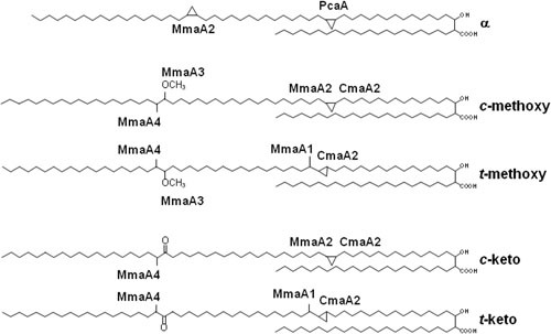
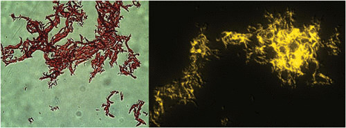
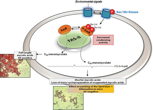
Acid-fast (AF) staining, also known as Ziehl-Neelsen stain microscopic detection, developed over a century ago, is even today the most widely used diagnostic method for tuberculosis. Herein we present a short historical review of the evolution of AF staining methods and discuss Koch's paradox, in which non-AF tubercle bacilli can be detected in tuberculosis patients or in experimentally infected animals. The conversion of Mycobacterium tuberculosis from an actively growing, AF-positive form to a nonreplicating, AF-negative form during the course of infection is now well documented. The mechanisms of loss of acid-fastness are not fully understood but involve important metabolic processes, such as the accumulation of triacylglycerol-containing intracellular inclusions and changes in the composition and spatial architecture of the cell wall. Although the precise component(s) responsible for the AF staining method remains largely unknown, analysis of a series of genetically defined M. tuberculosis mutants, which are attenuated in mice, pointed to the primary role of mycolic acids and other cell wall-associated (glyco)lipids as molecular markers responsible for the AF property of mycobacteria. Further studies are now required to better describe the cell wall reorganization that occurs during dormancy and to develop new staining procedures that are not affected by such cell wall alterations and that are capable of detecting AF-negative cells.


Figures

FIGURE 1
FIGURE 1
Chemical structures of the major mycolic acids of M. tuberculosis. Cyclopropane rings and methyl branches are shown and annotated with the S-adenosyl methionine-dependent methyl transferases responsible for their synthesis. c, cis; t, trans.
FIGURE 2
FIGURE 2
Staining of M. tuberculosis using ZN (left) and auramine O (right). Magnification, ×100.
FIGURE 3
FIGURE 3
Ser/Thr kinase-dependent signaling cascade resulting in phosphorylation of KasB and loss of acid-fastness. Modification of the cell wall composition in response to exogenous cues is central for M. tuberculosis adaptation to different environmental conditions. In response to an external signal, mycobacterial Ser/Thr kinases phosphorylate the different FAS-II components, including the β-ketoacyl ACP synthase KasB involved in the addition of the last carbon atoms during the mycolic acid elongation step. Phosphorylation on Thr334 and Thr336 decreases the condensation activity of KasB, resulting in the production of shorter mycolic acids, which probably affects the packing of the lipid layer and also results in the loss of the AF property and severe attenuation in mice.
FIGURE 4
FIGURE 4
Loss of AF staining coincides with the accumulation of TAG-containing intracellular lipid inclusions. (A) Dual staining of M. tuberculosis grown under multiple stress conditions, using auramine O for AF-staining (green) and Nile red as a neutral lipid stain (red). Bacilli were observed by confocal laser scanning microscopy. Overlaid images of the dual-stained bacteria are shown. Bar = 4 μm. (B) Quantification of the number of AF-positive and lipid-stain-positive bacilli grown as in (A). Auramine O-stained and Nile red-stained positive cells were counted from multiple scans. (Adapted from Deb et al. PLoS ONE 4(6):e6077 with permission of the publisher.)

Similar articles

Cited by

References

    1. Pawełczyk J, Kremer L. 2014. The molecular genetics of mycolic acid biosynthesis. Microbiol Spectr 2:MGM2–0003–2013. 10.1128/microbiolspec.MGM2-0003-2013. [PubMed] - DOI - PubMed
    1. Brennan PJ, Nikaido H. 1995. The envelope of mycobacteria. Annu Rev Biochem 64:29–63. 10.1146/annurev.bi.64.070195.000333 [PubMed] - DOI - PubMed
    1. Hoffmann C, Leis A, Niederweis M, Plitzko JM, Engelhardt H. 2008. Disclosure of the mycobacterial outer membrane: cryo-electron tomography and vitreous sections reveal the lipid bilayer structure. Proc Natl Acad Sci USA 105:3963–3967. 10.1073/pnas.0709530105 - DOI - PMC - PubMed
    1. Zuber B, Chami M, Houssin C, Dubochet J, Griffiths G, Daffé M. 2008. Direct visualization of the outer membrane of mycobacteria and corynebacteria in their native state. J Bacteriol 190:5672–5680. 10.1128/JB.01919-07 [PubMed] - DOI - PMC - PubMed
    1. Foulds J, O’Brien R. 1998. New tools for the diagnosis of tuberculosis: the perspective of developing countries. Int J Tuberc Lung Dis 2:778–783. [PubMed] - PubMed

MeSH terms